Category:Corpus Luteum
These pages and media relate to the specialized ovary structure the corpus luteum. Required for hormonal support of early pregnancy.
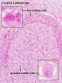
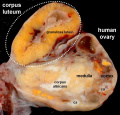
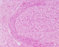
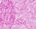
The corpus luteum is formed by both granulosa cells and thecal cells after ovulation has occurred. The wall of the follicle collapses into a folded structure, which is characteristic for the corpus luteum. Vascularization increases and a connective tissue network is formed. Theca interna cells and granulosa cells triple in size and start accumulating lutein within a few hours after ovulation. They are now called granulosa lutein cells and theca lutein cells and produce progesterone and oestrogens.
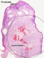
Hormone secretion in the corpus luteum ceases within 14 days after ovulation if the oocyte is not fertilised. In this case, the corpus luteum degenerates into a corpus albicans, a whitish scar tissue within the ovaries.
Pages in category 'Corpus Luteum'
The following 40 pages are in this category, out of 40 total.
C
P
- Paper - A study of the structure and vascular conditions of the human corpus luteum in the menstrual cycle and in pregnancy
- Paper - Cyclic changes in the ovaries and uterus of swine and their relations to the mechanism of implantation (1921)
- Paper - Mechanisms controlling the formation and persistence of the corpus luteum
- Paper - On the structure of the corpus luteum (1839)
- Paper - Studies on the human corpus luteum 1
- Paper - Studies on the human corpus luteum 2
- Paper - The corpus luteum in the ovary of the chicken (1918)
- Paper - The corpus luteum of pregnancy, as it is in swine (1915)
- Paper - The early development of the corpus luteum in the mare (1946)
- Paper - The events of the primate ovarian cycle
- Paper - The hormone of the corpus luteum
- Paper - The origin of the lutein cells of the corpus luteum
- Paper - The origin, growth and fate of the corpus luteum as observed in the ovary of the pig and man
- Template:Progesterone
R
- Template:Ref-AdamsHertig1969a
- Template:Ref-AdamsHertig1969b
- Template:Ref-AndersonMelampy1965
- Template:Ref-Clark1899
- Template:Ref-Corner1915
- Template:Ref-Corner1919
- Template:Ref-Corner1937
- Template:Ref-Corner1937b
- Template:Ref-Corner1952
- Template:Ref-Dalton1851
- Template:Ref-Falkiner1933
- Template:Ref-Harrison1946
- Template:Ref-Lee1839
- Template:Ref-Ninian1933
- Template:Ref-PearlBoring1918
- Template:Ref-Shaw1926
- Template:Ref-Solomons1924
Media in category 'Corpus Luteum'
The following 14 files are in this category, out of 14 total.
 Corner-plate01.jpg 653 × 935; 160 KB
 Corpus luteum lutein cells.jpg 450 × 600; 104 KB
 Corpus luteum.jpg 450 × 600; 94 KB
 Human ovary - corpus luteum 01.jpg 1,024 × 979; 162 KB
 Human ovary - corpus luteum 02.jpg 837 × 800; 119 KB
 Human ovary - corpus luteum 11.jpg 1,024 × 979; 89 KB
 Human ovary - corpus luteum 21.jpg 1,024 × 979; 89 KB
 Lutein cell glycogen granule em01.jpg 1,149 × 749; 169 KB
 Lutein cell lipid and glycogen em01.jpg 1,156 × 828; 149 KB
 Lutein cell lipid and glycogen em02.jpg 1,109 × 796; 227 KB
 Ovary corpus luteum.jpg 2,178 × 1,137; 376 KB
 Ovary histology 001.jpg 1,280 × 1,024; 360 KB
 Ovary histology 002.jpg 1,280 × 1,024; 270 KB
 Ovary histology 004.jpg 1,280 × 1,024; 401 KB


