File:Mouse zygote pronuclei 02.jpg
Original file (1,000 × 501 pixels, file size: 84 KB, MIME type: image/jpeg)
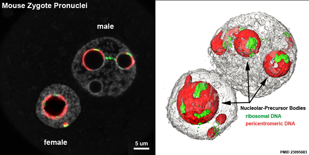
Mouse Zygote Pronuclei
Distribution of the pericentromeres, centromeres, and rDNA FISH signals in late 1-cell stage embryos.
1-cell embryos at the PN4 stage (collected at 27 hphCG) were processed with pericentromeric probes (red) and either rDNA probes (green, lower panel D/E/F).
DNA was counterstained with Yopro-1 (grey). Upper panel: (A/B) In both PNs, pericentromeres form more or less complete shells around the NPBs, in which the centromeres are embedded. Pericentromeres are also found at the nuclear periphery, associated with centromeric signals (see enlargement of A), and can form filaments with a “beads on a string” appearance (see enlargement of B). (C) 3D reconstruction of the same nuclei. Lower panel: (D, E) Most of the rDNA signals are around the NPBs. However, there are occasionally some signals associated with pericentromeric filaments (extending from the NPBs towards the nuclear periphery) as well as rDNA signals joining two NPBs. (F) 3D reconstruction of E.
Bar = 5 μm.
Reference
<pubmed>23095683</pubmed>| BMC Dev Biol.
Copyright
© 2012 Aguirre Lavin et al.; licensee BioMed Central Ltd. This is an Open Access article distributed under the terms of the Creative Commons Attribution License ( http://creativecommons.org/licenses/by/2.0), which permits unrestricted use, distribution, and reproduction in any medium, provided the original work is properly cited.
Figure 1. Panel E and F cropped, resized and relabelled.
File history
Click on a date/time to view the file as it appeared at that time.
| Date/Time | Thumbnail | Dimensions | User | Comment | |
|---|---|---|---|---|---|
| current | 10:43, 29 December 2012 | | 1,000 × 501 (84 KB) | Z8600021 (talk | contribs) |
You cannot overwrite this file.
File usage
The following page uses this file: