File:Melanoblast migration.png
From Embryology
No higher resolution available.
Melanoblast_migration.png (600 × 210 pixels, file size: 40 KB, MIME type: image/png)
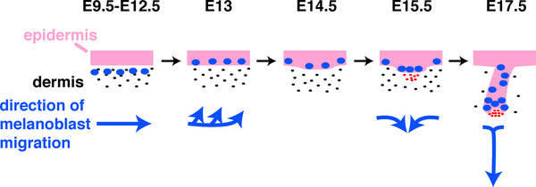
Figure 2. Schematic Depiction of Directions of Melanoblast Migration in Embryonic Mouse Skin from E9.5 to E17.5
Journal.pbio.0030372.g002.png
Pink, epithelium; black dots, dermal fibroblasts; blue ovals, melanoblasts; red dots, dermal condensate/DP.
File history
Click on a date/time to view the file as it appeared at that time.
| Date/Time | Thumbnail | Dimensions | User | Comment | |
|---|---|---|---|---|---|
| current | 08:17, 29 September 2009 | 600 × 210 (40 KB) | S8600021 (talk | contribs) | Figure 2. Schematic Depiction of Directions of Melanoblast Migration in Embryonic Mouse Skin from E9.5 to E17.5 Journal.pbio.0030372.g002.png Pink, epithelium; black dots, dermal fibroblasts; blue ovals, melanoblasts; red dots, dermal condensate/DP. |
You cannot overwrite this file.