Category:Electron Micrograph
From Embryology
This Embryology category shows pages and media related to the research imaging technique of scanning electron micrographs (SEM) in development. Note:
- images in this category may also include some of the associated bright field images taken before SEM fixation and imaging.
- there is a separate category Scanning Electron Micrograph.
- Links: Scanning Electron Microscopy
Pages in category 'Electron Micrograph'
The following 22 pages are in this category, out of 22 total.
H
P
- Paper - Cell-to-cell communication and ovulation - A study of the cumulus-oocyte complex
- Paper - Electron microscopy of the sperm tail - results obtained with a new fixative
- Paper - Fine structure of the human ovum in the pronuclear stage
- Paper - Studies on the fine structure of the mammalian testis 1
- Paper - Studies on the human oocyte and its follicle 1
- Template:Placenta EM links
R
Media in category 'Electron Micrograph'
The following file is in this category, out of 201 total.
(previous page) (next page)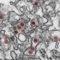 Zika virus TEM02.jpg 600 × 601; 128 KB
