File:Cullen1916 fig08.jpg
Original file (1,280 × 1,113 pixels, file size: 529 KB, MIME type: image/jpeg)
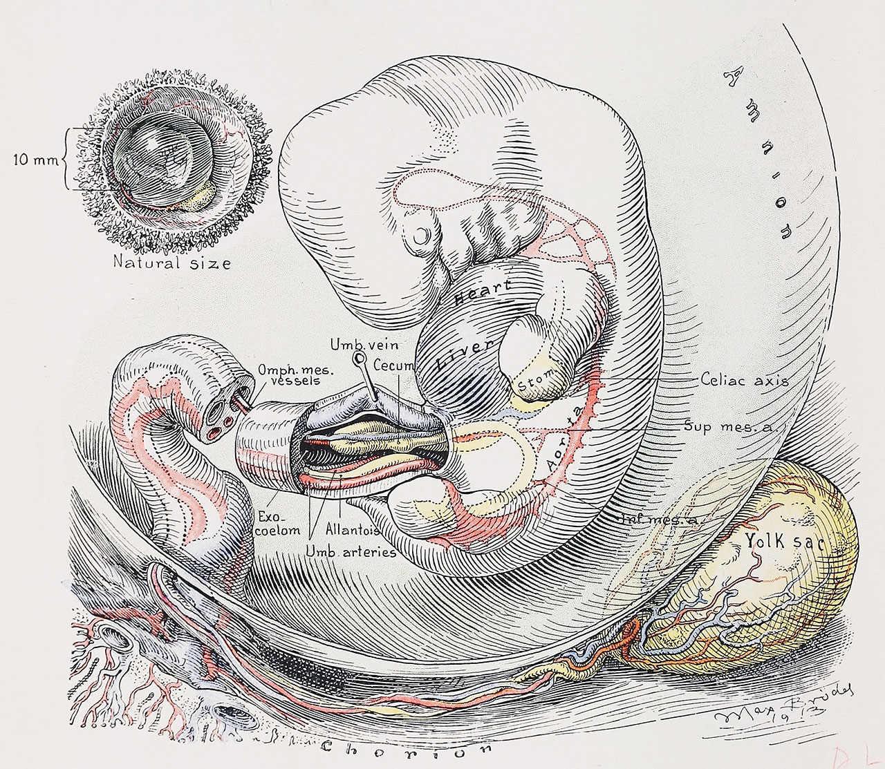
Fig. 8. Sagittal View of the Umbilical Region of a Human Embryo 10 mm in Length
The yolk-sac now lies far removed from the umbilical cord; its vessels and stalk follow a tortuous course. The umbilical cord here shows the first indication of a twist, which may be caused by the embryo rotating toward the right. In the embryos examined by us there has been but one exception to this rule. The small intestine extends a considerable distance into the exocoelom of the cord. At the outer end of the intestinal loop is situated the delicate omphalomesenteric duct. The omphalomesenteric vein passes on the left side of the intestinal loop; the artery, on the right side.
Reference
Cullen TS. Embryology, anatomy, and diseases of the umbilicus together with diseases of the urachus. (1916) W. B. Saunders Company, Philadelphia And London.
Cite this page: Hill, M.A. (2024, June 3) Embryology Cullen1916 fig08.jpg. Retrieved from https://embryology.med.unsw.edu.au/embryology/index.php/File:Cullen1916_fig08.jpg
- © Dr Mark Hill 2024, UNSW Embryology ISBN: 978 0 7334 2609 4 - UNSW CRICOS Provider Code No. 00098G
File history
Click on a date/time to view the file as it appeared at that time.
| Date/Time | Thumbnail | Dimensions | User | Comment | |
|---|---|---|---|---|---|
| current | 16:59, 27 October 2018 | | 1,280 × 1,113 (529 KB) | Z8600021 (talk | contribs) | |
| 16:58, 27 October 2018 | 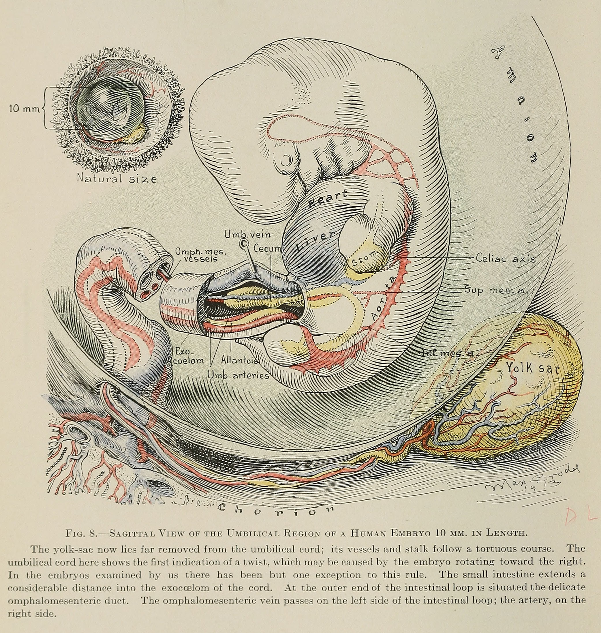 | 2,084 × 2,194 (1.2 MB) | Z8600021 (talk | contribs) | ===Reference=== {{Ref-Cullen1916}} {{Footer}} |
You cannot overwrite this file.
File usage
The following 3 pages use this file: