2010 Lab 3
Trilaminar Embryo to Early Embryo
Introduction
The first laboratories covered gametogenesis, fertilization, implantation and early events of embryonic development. The third week (Clinical week 5 LMP) is the begining of the embryonic period which continues through to week eight.
This laboratory will look at the third to fourth week of human development (stage 7 onward). We will be using a range of online materials and resources to explore these topics. The laboratory will also allow time for work on the group online project. Note that during this time the conceptus cells not contributing to the embryo are contributing to placental membranes and the early placenta.
The best way of beginning to understand the sequence is by considering the "stages" of development in relation to age (in days or weeks). Stages are based on the external and/or internal morphological development of the vertebrate embryo, and are not directly dependent on either age or size. Historically, the human embryonic period proper is divided into 23 Carnegie stages and criteria beyond morphological features include age in days, number of somites present, and embryonic length have sometimes been included in the stage descriptions. These stages are also shown in the movie Carnegie stage human growth through embryonic period.
Objectives
- Understand the conceptus changes during week 3 following implantation.
- Understand the process of gastrulation and the trilaminar embryo
- Understand the development of the germ layers: mesoderm and ectoderm
- Identify the early stages of embryonic development
Week 3
Folding
Endoderm, mesoderm and ectoderm layers. There are two major folding processes that take place during this time.
- Folding of the whole embryonic disc ventrally, separates the endoderm to form the epithelial lining of the gut. Folding of the embryonic disc occurs ventrally around the notochord, which forms a rod-like region running rostro-caudally in the midline.
- Folding of the ecoderm will form a neural groove, then closing to form a neural tube, separating the neural ectoderm from the embryo surface ectoderm.
Mesoderm Segmentation
Different regions of mesoderm form early intermediate structures.
- Somitogenesis - when part of the mesoderm layer segments during week 3 to form balls of mesoderm called somites. The later migration of cells forms the mesoderm germ layer. An embryonic connective tissue (mesenchyme) which forms nearly all the connective tissues of the body (the head is different). Somitogenesis is when part of this layer segments during week 3 to form balls of mesoderm called somites.
- Intraembryonic coelom - Within the embryonic disc lateral plate mesoderm a space (coelom) forms, it lies within the embryo and so is called the intraembryonic coelom. This single "horseshoe-shaped" space will form the 3 major body cavities: pericardial (around the heart), pleural (around the lungs) and peritoneal (around the GIT and visceral organs).
Ectoderm Segmentation
The central portion of the embryonic disc forms the neural plate, the edge of this plate forms neural crest and the edge forms the epitheium of the skin. This will be covered in week 4.
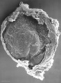
Stage 7
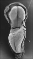
Stage 8
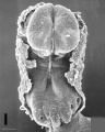
Stage 9


Folding
There are two major folding processes that take place during this time.
- Folding of the ecoderm will form a neural groove, then closing to form a neural tube, separating the neural ectoderm from the embryo surface ectoderm.
- Folding of the whole embryonic disc ventrally, separates the endoderm to form the epithelial lining of the gut. Folding of the embryonic disc occurs ventrally around the notochord, which forms a rod-like region running rostro-caudally in the midline.
![]()
![]()
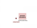
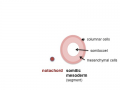
In relation to the notochord:
- Laterally (either side of the notochord) lies mesoderm.
- Rostrally (above the notochord end) lies the buccopharyngeal membrane, above this again is the mesoderm region forming the heart.
- Caudally (below the notochord end) lies the primitive streak (where gastrulation occurred), below this again is the cloacal membrane.
- Dorsally (above the notochord) lies the neural tube then ectoderm.
- Ventrally (beneath the notochord) lies the mesoderm then endoderm.
The ventral endoderm (shown yellow) has grown to line a space called the yolk sac. Folding of the embryonic disc "pinches off" part of this yolk sac forming the first primative GIT.

Mesoderm
Mesoderm means the "middle layer" and it is from this layer that nearly all the bodies connective tissues are derived. In early mesoderm development a number of transient structures will form and then be lost as tissue structure is patterned and organised. Humans are vertebrates, with a "backbone", and the first mesoderm structure we will see form after the notochord will be somites.
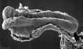
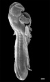
Facts: Week 4, 22 - 23 days, 2 - 3.5 mm, Somite Number 4 - 12
View: This is a dorsal view of the human embryo, the amniotic membrane has been removed. Top embryo is an early stage 10, bottom is late stage 10.
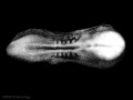
Early stage 10
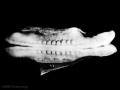
Late stage 10
Labeled stage 10


trilaminar embryo
mesoderm regions
somite coelom
neural tube and neural crest


Mesoderm Development
- epiblast -> mesoderm + axial mesoderm (notochord)
- lateral plate + paraxial mesoderm + axial mesoderm
- lateral plate + intermediate mesoderm + somites (body), paraxial mesoderm (head) + axial mesoderm
- somatic mesoderm + intraembryonic coelom + splanchnic mesoderm + intermediate mesoderm + somites (body), paraxial mesoderm (head) + axial mesoderm
Axial Mesoderm

 |
The notochord
Adult - contributes to the nucleus pulposus of the intervertebral disc |
Paraxial Mesoderm
 |
Adult - contributes vertebral column (vertebra and IVD), dermis of the skin, skeletal muscle of body and limbs |
Intermediate Mesoderm
 |
Adult - metanephros forms the kidney |
Lateral Plate Mesoderm
 |
Adult - body and limb connective tissues, gastrointestinal tract (connective tissues, muscle, organs), heart |
Somite Development

Somite initially forms 2 main components
- ventromedial- sclerotome forms vertebral body and intervertebral disc
- dorsolateral - dermomyotome forms dermis and skeletal muscle
paraxial mesoderm
early somite


Sclerotome
|
Myotome
 |
Forms 2 muscle groups in body and limbs
|
| Development of the sclerotome and myotome components of the somite. |
Dermatome
- connective tissue underlying epidermis
- begins as a dorsal thickening
- spreads throughout the body
Note - Dermatome is the term also used clinically postnatally to describe the region of skin supplied by a single spinal nerve.
Week 2 and 3 Movies
| Implantation | Mesoderm | Chorionic Cavity | Amniotic Cavity | Week 3 |
Embryo Stages and Events
| Day | Stage | Event |
| Stage 7 |  | |
| Stage 8 |  | |
 | ||
| Stage 9 | 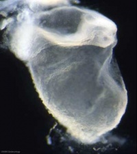 Musculoskeletal System Development somitogenesis - first somites form and continue to be added in sequence caudally Musculoskeletal System Development somitogenesis - first somites form and continue to be added in sequence caudally
Neural System Development - three main divisions of the brain, which are not cerebral vesicles, can be distinguished while the neural groove is still completely open
| |
| Cardiovascular System Development cardiogenesis - week 3 begins as paired heart tubes. |
Week 4
Key Events of Human Development during the fourth week (week 4) following fertilization or Clinical week 6 (LMP).
These notes cover the fourth week of embryonic development, which is the beginning of organogenesis, (specific tissues and systems are beginning to differentiate) from the trilaminar embryo. With many parallel processes, descriptions begin to get complicated! Many of the described processes begin and extend over a broader range of time. Some developmental processes will be discussed later in the practical to simplify matters.
Ectoderm on the embryo surface undergoes segmentation: The central portion of the embryonic disc forms the neural plate, the edge of this plate forms neural crest and the remainder outside this forms the epitheium of the skin and other structures.
Neurogenesis
- Central Nervous System (CNS) - the neural plate undergoes morphological changes to form the primitive central nervous system (brain, spinal cord). An epithelial layer of cells which contributes all neural (brain, spinal cord, peripheral nervous system) and the external epithelium (surface layer of the skin) of the embryo. Neurogenesis begins towards the end of week 3, when the neural tissues separate from this germ cell layer.
- Peripheral Nervous System (PNS) - the neural crest cells in the body region migrate and spread to different regions of the embryo forming the PNS (dorsal root ganglia, sympathetic ganglia, enteric nervous system) and many other embryonic tissues. Neural crest cells in the head region form skeletal and other structures.
Pharyngeal Arches
In the head region, a series of ventral folds form under the brain in a rostral to caudal sequence, these are the pharyngeal arches.
Placodes
In the head region, ectoderm small patches form pairs of specialised placodes that eventually contribute to specific sensory components, cranial ganglia and the anterior pituitary (adenohypophysis).
Limb Buds
In the body region, limb buds form initially as ectoderm and mesoderm (somitic and somatic) components and are the "paddle-like" projections from the trunk which will form all the upper and lower limb components. (An overview of limb development will be covered in week 8).
Cardiogenesis
Within the embryo mesoderm, the heart tube and vascular development continues. Cardiogenesis will be covered in week 5, when septation begins.
Stage 10
Stage 11

Stage 12
Stage 13


Neurogenesis


Developmental sequence: neural plate -> (day 18-19) neural groove -> neural tube -> Central Nervous System (brain and spinal cord)
- Central Nervous System (CNS) - the neural plate undergoes morphological changes to form the primitive central nervous system. An epithelial layer of cells which contributes all neural (brain, spinal cord, peripheral nervous system) and the external epithelium (surface layer of the skin) of the embryo. Neurogenesis begins towards the end of week 3, when the neural tissues separate from this germ cell layer.
- Peripheral Nervous System (PNS) - the neural crest cells in the body region migrate and spread to different regions of the embryo forming the PNS and many other embryonic tissues. Neural crest cells in the head region form skeletal and other structures.
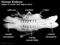
Human Neuralation - Early Stages
The stages below refer to specific Carneigie stages of development.
- stage 8 (about 18 postovulatory days) neural groove and folds are first seen
- stage 9 the three main divisions of the brain, which are not cerebral vesicles, can be distinguished while the neural groove is still completely open
- stage 10 (two days later) neural folds begin to fuse near the junction between brain and spinal cord, when neural crest cells are arising mainly from the neural ectoderm
- stage 11 (about 24 days) the cranial neuropore (rostral, cephalic or anterior) closes within a few hours; closure is bidirectional, it takes place from the dorsal and terminal lips and may occur in several areas simultaneously. The two lips, however, behave differently.
- stage 12 (about 26 days) The caudal (posterior) neuropore takes a day to close
- the level of final closure is approximately at future somitic pair 31
- corresponds to the level of sacral vertebra 2
- stage 13 (4 weeks) the neural tube is normally completely closed.
![]()
Three primary brain vesicles develop initially due to the neural plate being broader at the cranial (brain) end than the narrower caudal (spinal cord) end. When the plate fuses to form a tube, these 3 initial expansions (vesicles) result.

Pharyngeal Arches and Placodes
In the head region, two main components of head development form the pharyngeal arches and sensory placodes.
- Pharyngeal arches form a series of ventral folds under the brain in a rostral to caudal sequence. These arches will form most of the head and neck structures of the embryo and contain all three germ layers (ectoderm, mesoderm and endoderm). The topic of head and sensory development is covered in detail in BGD cycle B.
- Small patches of ectoderm form pairs of specialised placodes that eventually contribute to specific sensory components, cranial ganglia and the anterior pituitary (adenohypophysis).


Embryo Stage 13
Movies - Embryo Carnegie stage 13 - These are rotating animations based upon reconstruction of individual serial slice images of the stage 13 embryo.
| Central Nervous System | Gastrointestinal | Cardiovascular |
Week 4 Movies
Note that many of the movies start in week 4 and continue on through later embryonic development.
Ectoderm
| Neural Plate | Neural Tube |

|

|

|

|

|

|

|
Mesoderm
| Mesoderm | Somite Structures | Vertebra |
Endoderm
![]()
Neural Abnormalities


See also Neural Abnormalities
Neural Tube Defects (NTD)
- Failure of neural tube closure either incorrectly or incomplete.
- Dysraphism is the term often used to describe the defective fusion of the neural folds. The position and degree of failure of fusion will result in either embryonic death or a range of different neural defects. The way (mode) in which the human neural tube fuses has been a source of contention. In humans, fusion appears to initiate at multiple sites but the mode is different from that found in many animal models used in developmental studies. Severity dependent upon level within the tube and degree of failure (caudal - spina bifida; cranial - anancephaly)
Maternal Diet - Folate
Research demonstrated that that supplementation of maternal diet with folate reduces incidence of NTDs (More? Abnormal Development - Folic Acid and Neural Tube Defects)
UK
A randomised controlled trial conducted by the Medical Research Council of the United Kingdom demonstrated a 72% reduction in risk of recurrence by periconceptional (ie before and after conception) folic acid supplementation (4mg daily).
USA
Women who have one infant with a neural tube defect have a significantly increased risk of recurrence (40-50 per thousand compared with 2 per thousand for all births)


- Food and Drug Administration (USA) in 1996 authorized that all enriched cereal grain products be fortified with folic acid, with optional fortification beginning in March 1996 and mandatory fortification in January 1998. The data in the above graphs show the subsequent changes in anencephaly and spina bifida rate over that period.
Australia
- NHMRC policy statement (1993) emphasises the need for women who are capable of getting pregnant, or who are planning a pregnancy to be advised about folate and the importance of increasing folate intake to 0.4 - 0.5mg daily.
- Food Standards (FSANZ) had allowed industry two years to prepare to add folic acid to wheat flour used in making bread. Wheat flour will contain folic acid by 13 September 2009.
Links: Victoria - Folate information for health professionals | NHMRC - Nutrient Reference Values for Australia and New Zealand Including Recommended Dietary Intakes | NHMRC - Iodine supplementation for Pregnant and Breastfeeding Women
Embryo Stages and Events
| Day | Stage | Event |
| Stage 10 |  
Respiratory System Development
| |
| Heart begins to beat in humans by day 22-23, first functioning embryonic organ formed. | ||
| Stage 11 | 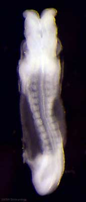
Thyroid thyroid median endodermal thickening in the floor of pharynx Neural System Development rostral (or cephalic) neuropore closes within a few hours; closure is bidirectional, it takes place from the dorsal and terminal lips and may occur in two areas simultaneously. The two lips, however, behave differently. Optic ventricle appears | |
| Stage 12 | 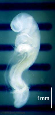
Pituitary Week 4 hypophysial pouch, Rathke’s pouch, diverticulum from roof GIT - Liver septum transversum forming liver stroma and hepatic diverticulum forming hepatic trabeculae PMID: 9407542 Neural System Development caudal neuropore takes a day to close (closure is approximately at future somitic pair 31/sacral vertebra 2) Neural System Development secondary neurulation begins Neural Crest Development cardiac crest, neural crest from rhombomeres 6 and 7 that migrates to pharyngeal arch 3 and from there the truncus arteriosus PMID: 17848161 Neural Crest Development vagal neural crest enter the foregut (20-25 somite stage) | |
| Stage 13 | 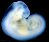 Neural System Development the neural tube is normally completely closed, ventricular system now separated from amniotic fluid. Neural crest at spinal level is segregating, and spinal ganglia are in series with the somites. Spinal cord ventral roots beginning to develop. PMID: 3354839 Neural System Development the neural tube is normally completely closed, ventricular system now separated from amniotic fluid. Neural crest at spinal level is segregating, and spinal ganglia are in series with the somites. Spinal cord ventral roots beginning to develop. PMID: 3354839
telencephalon cavity appears GIT - Liver epithelial cord proliferation enmeshing stromal capillaries PMID: 9407542 Sense - Smell Crest comes from the nasal platesPMID: 15604533 Skin 4 weeks - simple ectoderm epithelium over mesenchyme Skin 1-3 months ectoderm- germinative (basal) cell repeated division of generates stratified epithelium; mesoderm- differentiates into connective tissue and blood vessels |
Group Online Project
- After last weeks laboratory you should have begun researching your topic.
- Has everyone/anyone in your group tried to add something to the group discussion page?
- Have you found some relevant references and practiced making a reference link?
- Together as a group discuss the major concepts that should be included on the project page.
- Remember the Group Assessment Criteria
- Designate concept areas that each individual will begin researching/working on.
- Think about the mix of content (text/images). At this stage we are not concerned about the overall page layout or fine structure, this will come later after you have assembled the content you want to include.
- What will be the student contributed image on your page (you can have more than one) and who will do the work?
Glossary Links
- Glossary: A | B | C | D | E | F | G | H | I | J | K | L | M | N | O | P | Q | R | S | T | U | V | W | X | Y | Z | Numbers | Symbols | Term Link
Course Content 2010
Embryology Introduction | Cell Division/Fertilization | Lab 1 | Week 1&2 Development | Week 3 Development | Lab 2 | Mesoderm Development | Ectoderm, Early Neural, Neural Crest | Lab 3 | Early Vascular Development | Placenta | Lab 4 | Endoderm, Early Gastrointestinal | Respiratory Development | Lab 5 | Head Development | Neural Crest Development | Lab 6 | Musculoskeletal Development | Limb Development | Lab 7 | Kidney | Genital | Lab 8 | Sensory | Stem Cells | Stem Cells | Endocrine | Lab 10 | Late Vascular Development | Integumentary | Lab 11 | Birth, Postnatal | Revision | Lab 12 | Lecture Audio | Course Timetable
Cite this page: Hill, M.A. (2024, June 10) Embryology 2010 Lab 3. Retrieved from https://embryology.med.unsw.edu.au/embryology/index.php/2010_Lab_3
- © Dr Mark Hill 2024, UNSW Embryology ISBN: 978 0 7334 2609 4 - UNSW CRICOS Provider Code No. 00098G