File:Stage11 sem21.jpg

Stage11_sem21.jpg (600 × 447 pixels, file size: 79 KB, MIME type: image/jpeg)
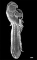
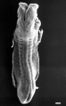
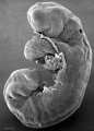
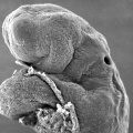
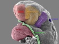
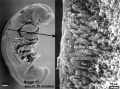
Human Embryo Carnegie stage 11
Carnegie stage 11, 25 days, 19 somite pairs, 100 μm scale bar.
Scanning EM lateral view embryo fractured dorsally (box region) to show the neural crest and neural tube.
Head Region - Cranial neuropore open. Otic placode indenting on dorsal side behind the second pharyngeal arch. First pharyngeal arch lying below and beside the stomedeum.
Body Region - Ventral body wall removed to show pericardial cavity and heart tube. Yolk sac also removed to show midgut.
Carnegie stage 11: Week 4, 23 - 26 days, 2.5 - 4.5 mm, Somite Number 13 - 20
- Stage 11 SEM Images: dorsolateral whole embryo | dorsal embryo | lateral embryo | lateral head | lateral head with overlay | embryo cross-section | ventrolateral head | ventrolateral head with overlay | ventral head | buccopharyngeal membrane | neural crest | posterior neuropore | anterior neuropore | Carnegie stage 11
- Human Embryo (stage 11)
dorsolateral whole embryo
dorsal embryo
lateral embryo
lateral head
lateral head with overlay

embryo cross-section
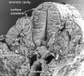
embryo cross-section label
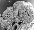
neural cross-section label

ventrolateral head
ventrolateral head with overlay

ventral head

buccopharyngeal membrane
neural crest
posterior neuropore
anterior neuropore


Image Source: Scanning electron micrographs of the Carnegie stages of the early human embryos are reproduced with the permission of Prof Kathy Sulik, from embryos collected by Dr. Vekemans and Tania Attié-Bitach. Images are for educational purposes only and cannot be reproduced electronically or in writing without permission.
- Carnegie Stages: 1 | 2 | 3 | 4 | 5 | 6 | 7 | 8 | 9 | 10 | 11 | 12 | 13 | 14 | 15 | 16 | 17 | 18 | 19 | 20 | 21 | 22 | 23 | About Stages | Timeline
Cite this page: Hill, M.A. (2024, May 23) Embryology Stage11 sem21.jpg. Retrieved from https://embryology.med.unsw.edu.au/embryology/index.php/File:Stage11_sem21.jpg
- © Dr Mark Hill 2024, UNSW Embryology ISBN: 978 0 7334 2609 4 - UNSW CRICOS Provider Code No. 00098G
File history
Click on a date/time to view the file as it appeared at that time.
| Date/Time | Thumbnail | Dimensions | User | Comment | |
|---|---|---|---|---|---|
| current | 16:03, 30 October 2010 | | 600 × 447 (79 KB) | S8600021 (talk | contribs) | '''Human Embryo''' Carnegie stage 11 25 days, 19 somite pairs Facts: Week 4, 23 - 26 days, 2.5 - 4.5 mm, Somite Number 13 - 20 View: This is a scanning EM of the embryo fractured to show the neural crest, neural tube, notochord and somites. Features: |
You cannot overwrite this file.
File usage
The following 60 pages use this file:
- Abnormal Development - Thalidomide
- BGDA Lecture - Development of the Nervous System
- BGDA Practical 7 - Week 4
- Carnegie stage 11
- Human Embryo SEM
- K12 Thalidomide
- Lecture - Ectoderm Development
- Lecture - Neural Crest Development
- Neural Crest - Cranial Nerve Development
- Neural Crest - Cranial Nerves
- Neural Crest - Enteric Nervous System
- Neural Crest - Peripheral Nervous System
- Neural Crest Development
- Science Student Projects
- Talk:Carnegie stage 11
- File:Stage11 sem10.jpg
- File:Stage11 sem100.jpg
- File:Stage11 sem100a.jpg
- File:Stage11 sem100b.jpg
- File:Stage11 sem100c.jpg
- File:Stage11 sem101.jpg
- File:Stage11 sem10a.jpg
- File:Stage11 sem10b.jpg
- File:Stage11 sem10c.jpg
- File:Stage11 sem13.jpg
- File:Stage11 sem13a.jpg
- File:Stage11 sem13b.jpg
- File:Stage11 sem13c.jpg
- File:Stage11 sem2.jpg
- File:Stage11 sem21.jpg
- File:Stage11 sem2a.jpg
- File:Stage11 sem2b.jpg
- File:Stage11 sem2c.jpg
- File:Stage11 sem3.jpg
- File:Stage11 sem3a.jpg
- File:Stage11 sem3b.gif
- File:Stage11 sem3b.jpg
- File:Stage11 sem3c.jpg
- File:Stage11 sem4.jpg
- File:Stage11 sem4a.jpg
- File:Stage11 sem4b.jpg
- File:Stage11 sem4c.jpg
- File:Stage11 sem5.jpg
- File:Stage11 sem5a.jpg
- File:Stage11 sem5b.jpg
- File:Stage11 sem5c.jpg
- File:Stage11 sem6.jpg
- File:Stage11 sem7.jpg
- File:Stage11 sem7a.jpg
- File:Stage11 sem7b.jpg
- File:Stage11 sem8.jpg
- File:Stage11 sem81.jpg
- File:Stage11 sem82.jpg
- File:Stage11 sem8a.jpg
- File:Stage11 sem8b.jpg
- File:Stage11 sem9.jpg
- File:Stage11 sem9a.jpg
- File:Stage11 sem9b.jpg
- Template:Carnegie stage 11-14 image table
- Template:Stage11SEM