File:Gray0610.jpg
Original file (303 × 1,000 pixels, file size: 81 KB, MIME type: image/jpeg)
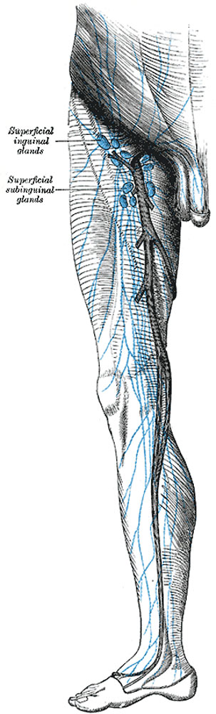
The superficial lymph glands and lymphatic vessels of the lower extremity
The inguinal glands (lymphoglandulæ inguinales) (Fig. 610), from twelve to twenty in number, are situated at the upper part of the femoral triangle. They may be divided into two groups by a horizontal line at the level of the termination of the great saphenous vein; those lying above this line are termed the superficial inguinal glands, and those below it the subinguinal glands, the latter group consisting of a superficial and a deep set.
Superficial Inguinal Glands form a chain immediately below the inguinal ligament. They receive as afferents lymphatic vessels from the integument of the penis, scrotum, perineum, buttock, and abdominal wall below the level of the umbilicus.
Superficial Subinguinal Glands (lymphoglandulæ subinguinales superficiales) are placed on either side of the upper part of the great saphenous vein; their efferents consist chiefly of the superficial lymphatic vessels of the lower extremity; but they also receive some of the vessels which drain the integument of the penis, scrotum, perineum, and buttock.
The Lymphatic Vessels of the Lower Extremity The lymphatic vessels of the lower extremity consist of two sets, superficial and deep, and in their distribution correspond closely with the veins.
The superficial lymphatic vessels lie in the superficial fascia, and are divisible into two groups: a medial, which follows the course of the great saphenous vein, and a lateral, which accompanies the small saphenous vein. The vessels of the medial group (Fig. 610) are larger and more numerous than those of the lateral group, and commence on the tibial side and dorsum of the foot; they ascend both in front of and behind the medial malleolus, run up the leg with the great saphenous vein, pass with it behind the medial condyle of the femur, and accompany it to the groin, where they end in the subinguinal group of superficial glands. The vessels of the lateral group arise from the fibular side of the foot; some ascend in front of the leg, and, just below the knee, cross the tibia to join the lymphatics on the medial side of the thigh; others pass behind the lateral malleolus, and, accompanying the small saphenous vein, enter the popliteal glands.
(Text from Gray's Anatomy 1918)
- Gray's Images: Development | Lymphatic | Neural | Vision | Hearing | Somatosensory | Integumentary | Respiratory | Gastrointestinal | Urogenital | Endocrine | Surface Anatomy | iBook | Historic Disclaimer
| Historic Disclaimer - information about historic embryology pages |
|---|
 |
| iBook - Gray's Embryology | |
|---|---|

|
|
Reference
Gray H. Anatomy of the human body. (1918) Philadelphia: Lea & Febiger.
Cite this page: Hill, M.A. (2024, May 23) Embryology Gray0610.jpg. Retrieved from https://embryology.med.unsw.edu.au/embryology/index.php/File:Gray0610.jpg
- © Dr Mark Hill 2024, UNSW Embryology ISBN: 978 0 7334 2609 4 - UNSW CRICOS Provider Code No. 00098G
File history
Click on a date/time to view the file as it appeared at that time.
| Date/Time | Thumbnail | Dimensions | User | Comment | |
|---|---|---|---|---|---|
| current | 22:54, 14 February 2013 | 303 × 1,000 (81 KB) | Z8600021 (talk | contribs) | (Text from Gray's Anatomy 1918) {{Gray Anatomy}} Category:Immune |
You cannot overwrite this file.
File usage
The following 3 pages use this file: