Uploads by MarkHill
From Embryology
This special page shows all uploaded files.
| Date | Name | Thumbnail | Size | Description | Versions |
|---|---|---|---|---|---|
| 12:04, 20 August 2009 | Weblog.png (file) |  |
2 KB | UNSW Embryology Weblog banner icon | 1 |
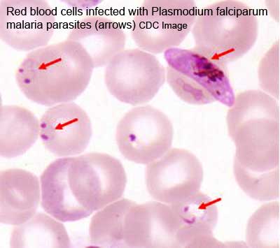
| 10:16, 18 August 2009 | Malaria plasmodium falciparum.jpg (file) |  |
14 KB | Malaria (plasmodium falciparum) Image source: CDC | 1 |
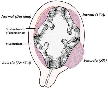
| 10:11, 18 August 2009 | Placenta abnormalities.jpg (file) |  |
37 KB | 1 | |
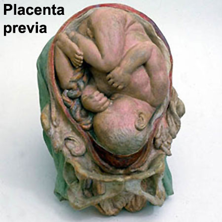
| 10:05, 18 August 2009 | Galletti1770 placenta previa.jpg (file) |  |
36 KB | 1 | |
| 17:08, 17 August 2009 | Gray0039.gif (file) |  |
43 KB | Scheme of placental circulation. | 1 |
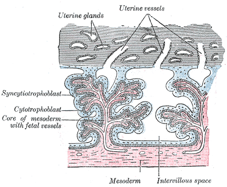
| 17:07, 17 August 2009 | Gray0037.gif (file) |  |
37 KB | Secondary chorionic villi. Diagrammatic. (Modified from Bryce.) | 1 |
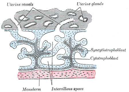
| 17:06, 17 August 2009 | Gray0036.gif (file) |  |
26 KB | Primary chorionic villi. Diagrammatic. (Modified from Bryce.) | 1 |
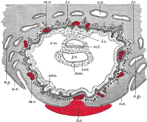
| 17:04, 17 August 2009 | Gray0032.gif (file) |  |
57 KB | Section through ovum imbedded in the uterine decidua. Semidiagrammatic. (After Peters.) * am. Amniotic cavity * b.c. Blood-clot * b.s. Body-stalk. * ect. Embryonic ectoderm * ent. Entoderm * mes. Mesoderm. * m.v. Maternal vessels. * tr. Trophoblast. | 1 |
| 14:42, 17 August 2009 | Gray0024.gif (file) |  |
5 KB | Diagram showing earliest observed stage of human ovum. | 1 |
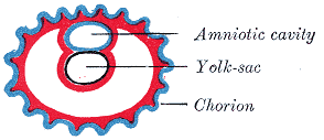
| 14:41, 17 August 2009 | Gray0025.gif (file) |  |
7 KB | Diagram illustrating early formation of allantois and differentiation of body-stalk. | 1 |
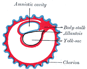
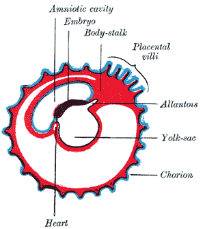
| 14:41, 17 August 2009 | Gray0026.gif (file) |  |
9 KB | Diagram showing later stage of allantoic development with commencing constriction of the yolk-sac. | 1 |
| 14:40, 17 August 2009 | Gray0027.gif (file) |  |
10 KB | Diagram showing the expansion of amnion and delimitation of the umbilicus. | 1 |
| 14:16, 12 August 2009 | Abnormal AusData81-92Graph.png (file) |  |
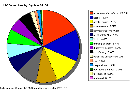
7 KB | Pie diagram shows the percentage of developmental abnormalities by categories of all notifiable birth defects in Australia. Data groupings and classification as Major or Minor Abnormalities are based on that used by the Australian Institute of Health and | 1 |
| 14:13, 12 August 2009 | Abnormal AusData81-92.png (file) |  |
10 KB | Pie diagram shows the percentage of developmental abnormalities by categories of all notifiable birth defects in Australia. Data groupings and classification as Major or Minor Abnormalities are based on that used by the Australian Institute of Health and | 1 |
| 11:58, 12 August 2009 | Spinal cord delta notch model.png (file) |  |
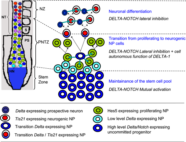
171 KB | Figure 9. A working model for the involvement of DELTA-NOTCH signalling in the transition from proliferation to neurogenesis in the developing chick spinal cord. Schematic model of the embryonic rostro-caudal gradient of neurogenesis along the prospectiv | 1 |
| 11:42, 12 August 2009 | Abnormal81-92-neuron.png (file) |  |
9 KB | Pie diagram shows the percentage of neural defects of all notifiable birth defects in Australia. Data groupings and classification as Major or Minor Abnormalities are based on that used by the Australian Institute of Health and Welfare National Perinatal | 1 |
| 01:07, 11 August 2009 | CNS later development.jpg (file) |  |
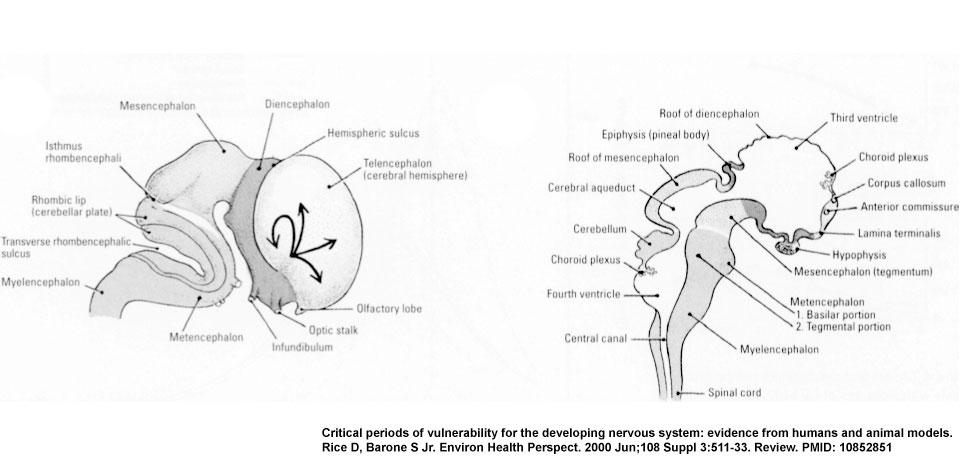
59 KB | CNS later development cartoon (E) The lateral view shows the migratory paths from the more central ventricular zone and gradients maturation of the neocortex (see arrows). (F) The midsagittal view of the brain and spinal cord, with the major divisions d | 1 |
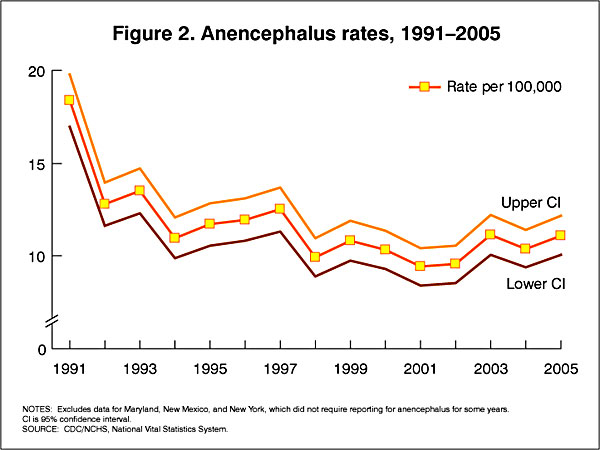
| 16:07, 10 August 2009 | USA anencephaly rates.jpg (file) |  |
45 KB | In the U.S.A. the Food and Drug Administration in 1996 authorized that all enriched cereal grain products be fortified with folic acid, with optional fortification beginning in March 1996 and mandatory fortification in January 1998. The data below shows t | 1 |
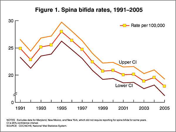
| 16:07, 10 August 2009 | USA spina bifida rates.jpg (file) |  |
46 KB | In the U.S.A. the Food and Drug Administration in 1996 authorized that all enriched cereal grain products be fortified with folic acid, with optional fortification beginning in March 1996 and mandatory fortification in January 1998. The data below shows t | 1 |
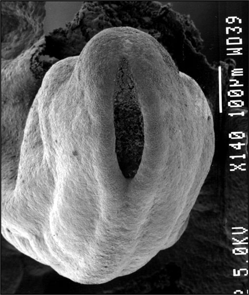
| 15:43, 10 August 2009 | Stage12 SEM3.jpg (file) |  |
68 KB | Original file name: Stage12semneuropore.jpg | 1 |
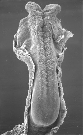
| 15:32, 10 August 2009 | Stage10 SEM1.jpg (file) |  |
28 KB | Carnegie Stages 10, 4-5 somites Features: brain fold, neural groove, amniotic sac, presomitic mesoderm, embryonic disc, primitive node, primative streak, primative groove, connecting stalk Facts: Week 3, 21 days, 4 - 5 somites, View: Dorsal view amnioti | 1 |
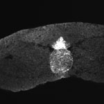
| 15:22, 10 August 2009 | Stage22 HPA2L.jpg (file) |  |
82 KB | Original File name: HUMHPA2L.GIF | 1 |
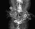
| 15:22, 10 August 2009 | Stage22 HPA1L.jpg (file) |  |
65 KB | Original file name: HUMHPA1L.GIF | 1 |
| 15:08, 10 August 2009 | Neural plate movie icon.jpg (file) | 4 KB | 1 | ||
| 15:07, 10 August 2009 | Csf cartoon3.jpg (file) |  |
37 KB | 1 | |
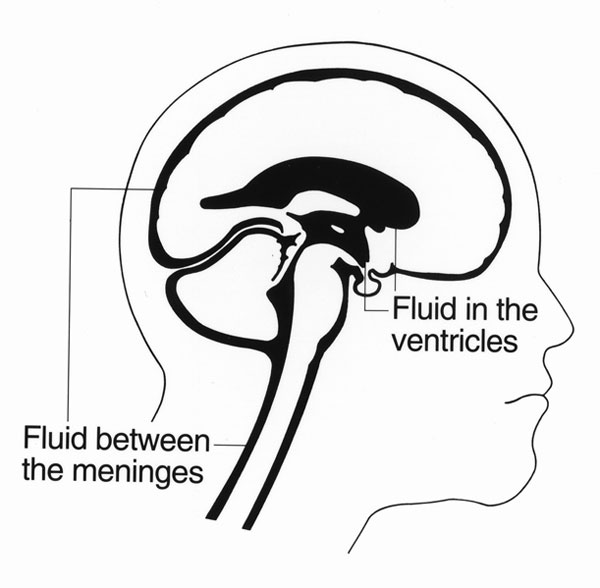
| 15:07, 10 August 2009 | Csf cartoon2.jpg (file) |  |
44 KB | Cerebrospinal fluid (CSF) location in the brain and spinal cord (CNS) - cartoon | 1 |
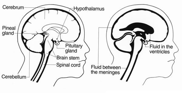
| 15:06, 10 August 2009 | Csf cartoon1.jpg (file) |  |
35 KB | Cerebrospinal fluid (CSF) location in the brain and spinal cord (CNS) - cartoon | 1 |
| 15:04, 10 August 2009 | Neural tube defect meningomyelocele.jpg (file) |  |
13 KB | Neural tube defect - meningomyelocele Source: UNSW Embryology | 1 |
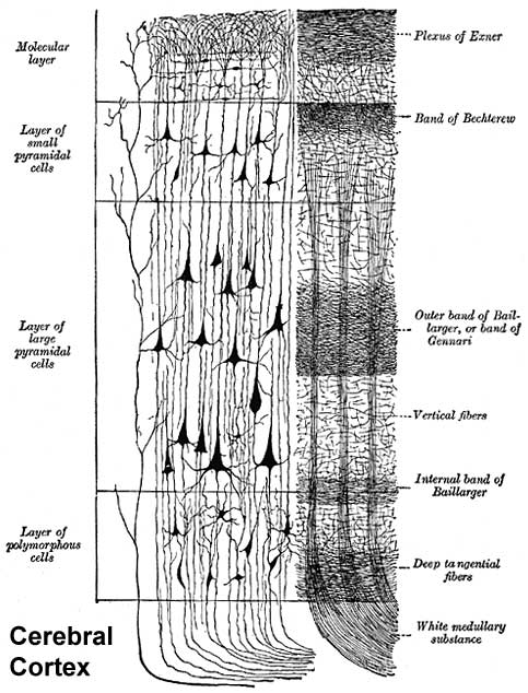
| 15:03, 10 August 2009 | Historic-Cerebral-cortex.jpg (file) |  |
65 KB | 1 | |
| 15:03, 10 August 2009 | Historic-Neural-plate.jpg (file) |  |
36 KB | Human Embryo neural plate historic drawing 2 mm embryo dorsal view | 1 |
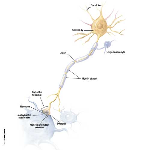
| 15:01, 10 August 2009 | Neuron cartoon.jpg (file) |  |
11 KB | Neuron cartoon Original File name: Neuron1.jpg Source: NIH USA | 1 |
| 15:00, 10 August 2009 | Sonic hedgehog expression.jpg (file) |  |
7 KB | 1 | |
| 14:54, 10 August 2009 | Neural-crest-icon.png (file) | 7 KB | 1 | ||
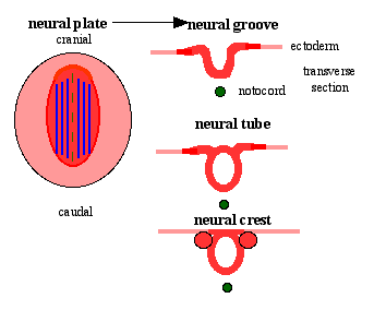
| 14:31, 10 August 2009 | Neuralplate cartoon.png (file) |  |
5 KB | Neural plate_cartoon | 1 |
| 14:28, 10 August 2009 | Stage10 neural sm.jpg (file) |  |
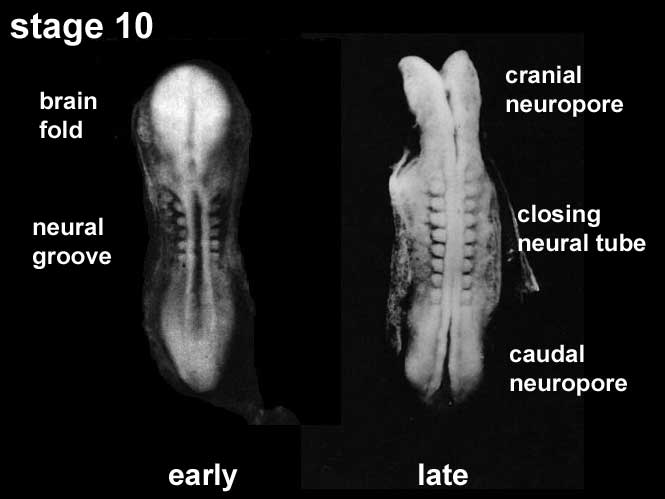
22 KB | Carnegie stage 10 small image showing neuralation About Carnegie stage 10 Facts: Week 4, 22 - 23 days, 2 - 3.5 mm, Somite Number 4 - 12 View: This is a dorsal view of the embryo. Top embryo is an early stage 10, bottom is late stage 10. Amniotic membra | 1 |
| 11:30, 10 August 2009 | Stage7 lateral-plate.jpg (file) |  |
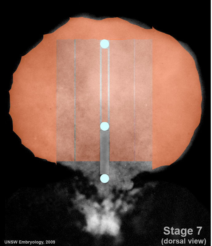
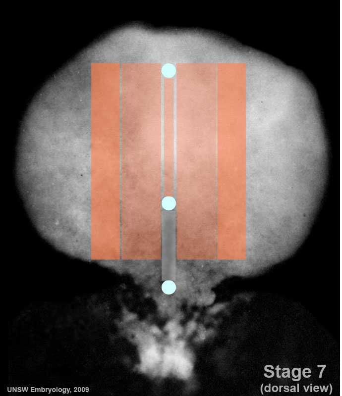
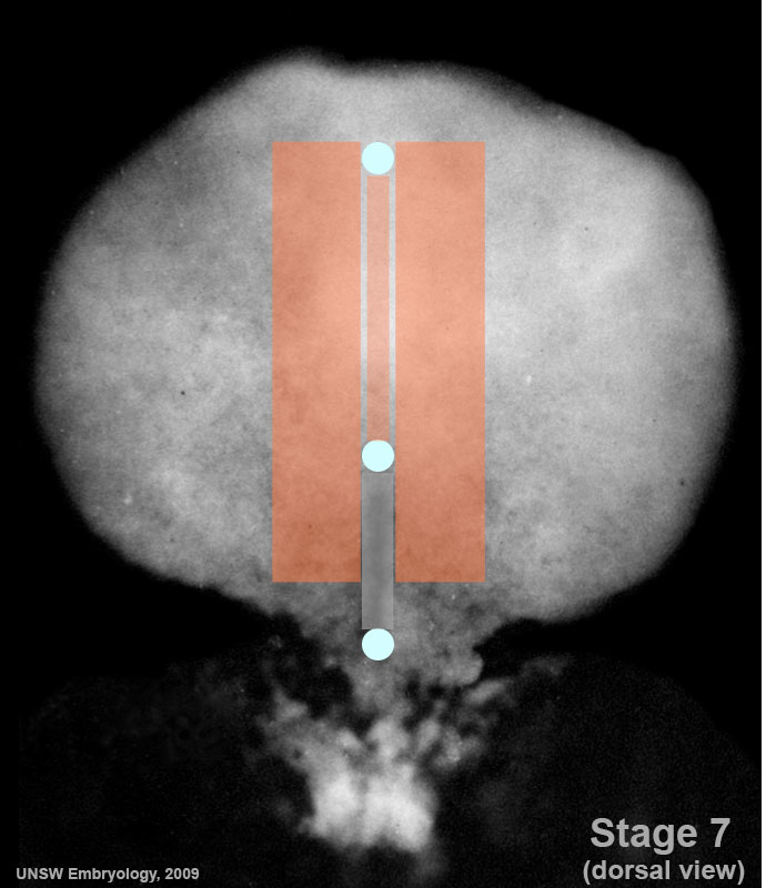
65 KB | Carnegie Stages 7 showing the lateral plate mesoderm region of the embryonic disc. Features: embryonic disc, primitive node, primative streak, primative groove, yolk sac Facts: Week 3, 15 - 17 days, 0.4 mm View 1: embryonic disc, showing the epiblast v | 1 |
| 11:29, 10 August 2009 | Stage7 intermediate-mesoderm.jpg (file) |  |
67 KB | Carnegie Stages 7 showing the intermediate mesoderm region of the embryonic disc. Features: embryonic disc, primitive node, primative streak, primative groove, yolk sac Facts: Week 3, 15 - 17 days, 0.4 mm View 1: embryonic disc, showing the epiblast vi | 1 |
| 11:29, 10 August 2009 | Stage7 paraxial-mesoderm.jpg (file) |  |
69 KB | Carnegie Stages 7 showing the paraxial mesoderm region of the embryonic disc. Features: embryonic disc, primitive node, primative streak, primative groove, yolk sac Facts: Week 3, 15 - 17 days, 0.4 mm View 1: embryonic disc, showing the epiblast viewed | 1 |
| 11:21, 10 August 2009 | Stage7 notochord.jpg (file) |  |
70 KB | Carnegie Stages 7 showing the notochord or axial mesoderm region of the embryonic disc. Features: embryonic disc, primitive node, primative streak, primative groove, yolk sac Facts: Week 3, 15 - 17 days, 0.4 mm View 1: embryonic disc, showing the epibl | 1 |
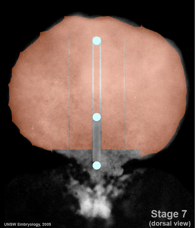
| 11:15, 10 August 2009 | Stage7 cloacal-oral-membranes.jpg (file) |  |
70 KB | Carnegie Stages 7 showing approximate regions where buccopharyngeal and cloacal membranes would lie on the embryonic disc. Features: embryonic disc, primitive node, primative streak, primative groove, yolk sac Facts: Week 3, 15 - 17 days, 0.4 mm View 1 | 1 |
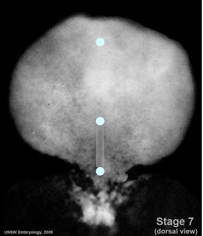
| 11:13, 10 August 2009 | Stage7 primitive-streak-node.jpg (file) |  |
69 KB | Carnegie Stages 7 showing the primitive streak and node regions of the embryonic dics Features: embryonic disc, primitive node, primative streak, primative groove, yolk sac Facts: Week 3, 15 - 17 days, 0.4 mm View 1: embryonic disc, showing the epiblas | 1 |
| 11:12, 10 August 2009 | Stage7 mesoderm.jpg (file) |  |
67 KB | Carnegie Stages 7 showing mesoderm region of embryonic disc Features: embryonic disc, primitive node, primative streak, primative groove, yolk sac Facts: Week 3, 15 - 17 days, 0.4 mm View 1: embryonic disc, showing the epiblast viewed from the amniotic | 1 |
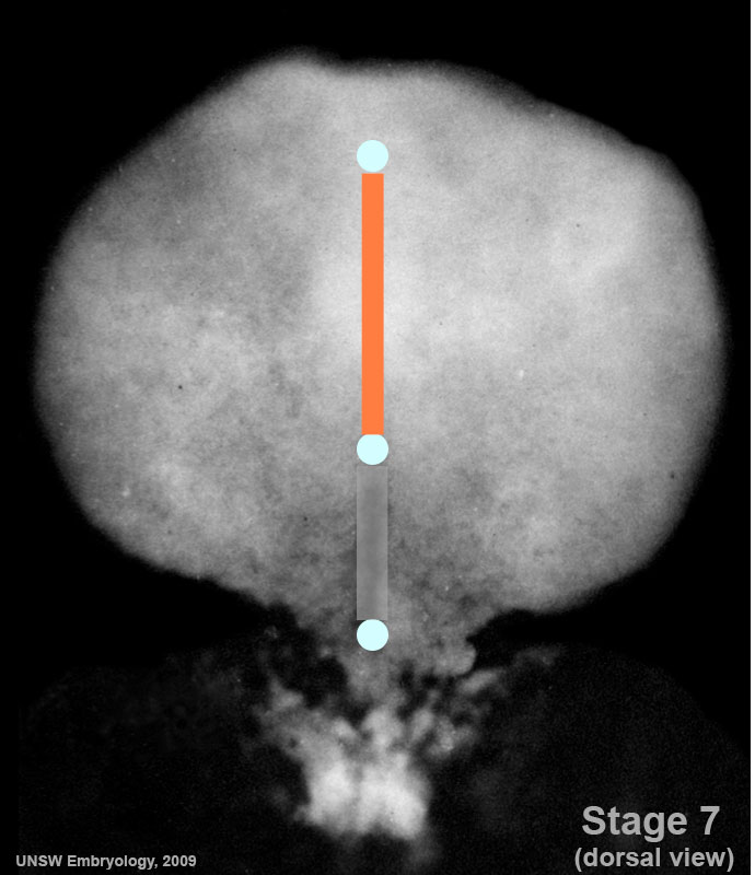
| 11:11, 10 August 2009 | Stage7 800x700px.jpg (file) |  |
69 KB | Carnegie Stages 7 Features: embryonic disc, primitive node, primative streak, primative groove, yolk sac Facts: Week 3, 15 - 17 days, 0.4 mm View 1: embryonic disc, showing the epiblast viewed from the amniotic (dorsal) side. Stage 7 Labelled | Stage | 1 |
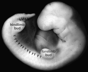
| 10:49, 10 August 2009 | Stage14 somites limbbuds.png (file) |  |
24 KB | Image source: UNSW Embryology http://embryology.med.unsw.edu.au/Notes/skmus.htm#Somite1 Category:Mesoderm Category:Somite | 1 |
| 15:37, 6 August 2009 | Connotea 32x32.png (file) |  |
1 KB | 1 | |
| 15:36, 6 August 2009 | Icon citeulike 16x16.gif (file) |  |
79 bytes | 1 | |
| 15:36, 6 August 2009 | Citeulike 16x16.png (file) |  |
413 bytes | 1 | |
| 15:35, 6 August 2009 | Delicious 32x32.png (file) |  |
888 bytes | 1 | |
| 15:35, 6 August 2009 | Reddit 32x32.png (file) |  |
2 KB | 1 | |
| 15:35, 6 August 2009 | Stumbleupon 32x32.png (file) |  |
2 KB | 1 |