File:Dog liver portosystemic shunts.jpg: Difference between revisions
(==Dog Liver PortoSystemic Shunts== Image shows the anatomy of a normal liver and of livers with intra- and extrahepatic portosystemic shunts. Congenital disorders of the hepatic portal vasculature are rare in man but occur frequently in certain dog breed) |
No edit summary |
||
| Line 1: | Line 1: | ||
==Dog Liver PortoSystemic Shunts== | ==Dog Liver PortoSystemic Shunts== | ||
Image shows the anatomy of dog normal liver and of livers with intra- and extrahepatic portosystemic shunts. Congenital disorders of the hepatic portal vasculature are rare in man but occur frequently in certain dog breeds. | |||
a No connection of blood vessels in the liver is seen within a normal liver resulting in a blood flow through the hepatic sinusoids. | '''a''' No connection of blood vessels in the liver is seen within a normal liver resulting in a blood flow through the hepatic sinusoids. | ||
b In case of PSS, blood bypasses the liver sinusoids and is therefore not subjected to hepatic metabolism. The intrahepatic shunt represents an abnormal connection of the portal vein with the systemic circulation, which is seen inside the liver. | '''b''' In case of PSS, blood bypasses the liver sinusoids and is therefore not subjected to hepatic metabolism. The intrahepatic shunt represents an abnormal connection of the portal vein with the systemic circulation, which is seen inside the liver. | ||
c In the case of an extrahepatic shunt, the aberrant connection is located outside the liver. | '''c''' In the case of an extrahepatic shunt, the aberrant connection is located outside the liver. | ||
===Reference=== | ===Reference=== | ||
<pubmed>22052005</pubmed>| [http://www.ncbi.nlm.nih.gov/pmc/articles/PMC3275728 PMC3275728] | [http://www.springerlink.com/content/yh45045r673r7561/ Mamm Genome.] | |||
This article is distributed under the terms of the Creative Commons Attribution Noncommercial License which permits any noncommercial use, distribution, and reproduction in any medium, provided the original author(s) and source are credited. | |||
[[Category:Dog]] [[Category:Liver]] [[Category:Cardiovascular]] [[Category:Cartoon]] | |||
Latest revision as of 08:12, 5 April 2012
Dog Liver PortoSystemic Shunts
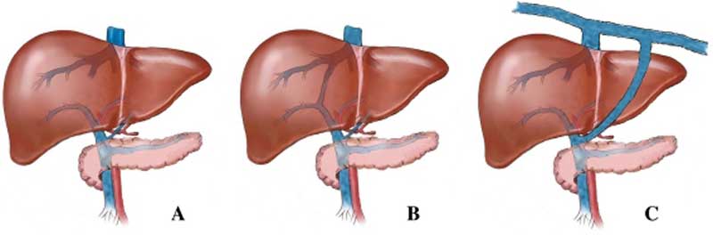
Image shows the anatomy of dog normal liver and of livers with intra- and extrahepatic portosystemic shunts. Congenital disorders of the hepatic portal vasculature are rare in man but occur frequently in certain dog breeds.
a No connection of blood vessels in the liver is seen within a normal liver resulting in a blood flow through the hepatic sinusoids.
b In case of PSS, blood bypasses the liver sinusoids and is therefore not subjected to hepatic metabolism. The intrahepatic shunt represents an abnormal connection of the portal vein with the systemic circulation, which is seen inside the liver.
c In the case of an extrahepatic shunt, the aberrant connection is located outside the liver.
Reference
<pubmed>22052005</pubmed>| PMC3275728 | Mamm Genome.
This article is distributed under the terms of the Creative Commons Attribution Noncommercial License which permits any noncommercial use, distribution, and reproduction in any medium, provided the original author(s) and source are credited.
File history
Click on a date/time to view the file as it appeared at that time.
| Date/Time | Thumbnail | Dimensions | User | Comment | |
|---|---|---|---|---|---|
| current | 08:08, 5 April 2012 | 800 × 264 (19 KB) | Z8600021 (talk | contribs) | ==Dog Liver PortoSystemic Shunts== Image shows the anatomy of a normal liver and of livers with intra- and extrahepatic portosystemic shunts. Congenital disorders of the hepatic portal vasculature are rare in man but occur frequently in certain dog breed |
You cannot overwrite this file.
File usage
The following page uses this file: