File:Stage 13 image 068.jpg: Difference between revisions
No edit summary |
No edit summary |
||
| Line 10: | Line 10: | ||
* '''Pleuroperitoneal fold''' on the medial side of the R common cardinal vein - this fold will form part of the diaphragm. | * '''Pleuroperitoneal fold''' on the medial side of the R common cardinal vein - this fold will form part of the diaphragm. | ||
:'''Links:''' [[Gastrointestinal Tract Development]] | [[Coelomic Cavity Development]] | [[Respiratory_System_-_Diaphragm|Diaphragm]] | :'''Links:''' [[Gastrointestinal Tract Development]] | [[Coelomic Cavity Development]] | [[Respiratory_System_-_Diaphragm|Diaphragm]] | ||
===Head and Neck=== | ===Head and Neck=== | ||
Latest revision as of 10:00, 9 June 2011
C5L Image Features
Gastrointestinal Tract
- Oesophagus is smaller while the trachea expands.
- Septum transversum ventral anchoring of attachment site is at the most cranial extension. This attachment now divides the intraembryonic coelom around the trachea into two canals, the L and R pleuro (pericardio-peritoneal) canals.
- Canals are lined by coelomic mesothelium and are continuous with whole I-E coelom (referred to hereafter simply as coelomic canals).
- Pleuroperitoneal fold on the medial side of the R common cardinal vein - this fold will form part of the diaphragm.
Head and Neck
Cardiovascular System
Cardiovascular System Development
Gastrointestinal System
Gastrointestinal Tract Development
Smaller oesophagus, expanding trachea. Note ventral anchoring of attachment site is at the most cranial extension of the septum transversum. Note also that this attachment now divides the intraembryonic coelom around the trachea into two canals, the L and R pleuro (pericardio-peritoneal) canals. (Canals are lined by coelomic mesothelium and are continuous with whole I-E coelom - they will be referred to hereafter simply as coelomic canals). Note the pleuroperitoneal fold on the medial side of the R common cardinal vein - this fold will form part of the diaphragm.
About Stage 13 Embryo Sections - This image is from a serial section of a 6mm CRL pig embryo with some features of the Stage 14 embryo. This embryo is approximately equal to the day 42 human embryo. Use these serial images to identify internal features and relationships that exist within the embryo at this stage. Then compare these images with the later features of the Carnegie stage 22 human embryo.
| Stage 13 Serial unlabeled images | Embryo Stage 13 Serial labeled images |
| System Links: Introduction | Cardiovascular | Coelomic Cavity | Endocrine | Gastrointestinal Tract | Genital | Head | Immune | Integumentary | Musculoskeletal | Neural | Neural Crest | Placenta | Renal | Respiratory | Sensory | Birth |
Cite this page: Hill, M.A. (2024, June 14) Embryology Stage 13 image 068.jpg. Retrieved from https://embryology.med.unsw.edu.au/embryology/index.php/File:Stage_13_image_068.jpg
- © Dr Mark Hill 2024, UNSW Embryology ISBN: 978 0 7334 2609 4 - UNSW CRICOS Provider Code No. 00098G
File history
Click on a date/time to view the file as it appeared at that time.
| Date/Time | Thumbnail | Dimensions | User | Comment | |
|---|---|---|---|---|---|
| current | 18:16, 10 August 2010 | 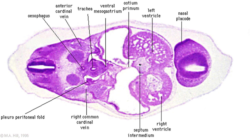 | 1,000 × 557 (97 KB) | S8600021 (talk | contribs) | {{Template:Stage13sections}} {{Template:Systems}} {{Template:Footer}} Category:Carnegie Stage 13 |
You cannot overwrite this file.
File usage
The following 20 pages use this file:
- 2010 Lab 5
- 2011 Lab 5 - Early Embryo
- ANAT2341 Lab 3 - Week 4
- ANAT2341 Lab 5 - Early Embryo
- BGDA Practical 7 - Week 5
- BGDB Gastrointestinal - Early Embryo
- Cardiovascular System - Developmental Shunts
- Carnegie stage 13
- Carnegie stage 13 - serial sections
- Embryo Serial Sections
- Fetal ECHO Meeting 2012
- Gastrointestinal Tract - Carnegie Stage 13
- Museum of Natural History Berlin - 2013 Seminar
- Neural 3D stage 13 Movie
- RPAH Cardiac Embryology 2014
- Respiratory System - Carnegie Stage 13
- Talk:Carnegie stage 13 - serial sections
- File:Stage14 respiratory tract.jpg
- Template:Stage13Licon120
- Template talk:Stage13Licon120