Category:Third Trimester: Difference between revisions
From Embryology
mNo edit summary |
mNo edit summary |
||
| Line 1: | Line 1: | ||
This {{Embryology}} category shows content related to the third trimester of human development. | This {{Embryology}} category shows content related to the {{third trimester}} of human development. | ||
Latest revision as of 10:26, 26 May 2020
This Embryology category shows content related to the third trimester of human development.
- Links: Second Trimester | Third Trimester | Fetal Development | Birth
| Links: human timeline | first trimester timeline | second trimester timeline | third trimester timeline | ||
| Event | ||
| Clinical third trimester |  hearing 3rd Trimester - vibration acoustically of maternal abdominal wall induces startle respone in fetus. hearing 3rd Trimester - vibration acoustically of maternal abdominal wall induces startle respone in fetus.
| |
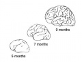
| respiratory Month 7 - respiratory bronchioles proliferate and end in alveolar ducts and sacs | ||
|
tooth Week 29 - Permanent premolars (correspond to the milk molars) appear. | ||
|
Genital male gonad (testes) descending | ||
| nail fingernails reach digit tip | ||
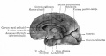
| neural brain cortical sulcation - primary sulci present[1] | ||
| neural brain cortical sulcation - insular, cingular, and occipital secondary sulci present[1] | ||
 Nail Development toenails reach digit tip Nail Development toenails reach digit tip
Lens Development - lens growth and interocular distance plateaus after 36 weeks of gestation[2] | ||
| Birth |  Clinical Week 40 Clinical Week 40
Heart pressure difference closes foramen ovale leaving a fossa ovalis thyroid TSH levels increase, thyroxine (T3) and T4 levels increase to 24 h, then 5-7 days postnatal decline to normal levels adrenal - zona glomerulosa, zona fasiculata present | |
- ↑ 1.0 1.1 Garel C, Chantrel E, Brisse H, Elmaleh M, Luton D, Oury JF, Sebag G & Hassan M. (2001). Fetal cerebral cortex: normal gestational landmarks identified using prenatal MR imaging. AJNR Am J Neuroradiol , 22, 184-9. PMID: 11158907
- ↑ Paquette LB, Jackson HA, Tavaré CJ, Miller DA & Panigrahy A. (2009). In utero eye development documented by fetal MR imaging. AJNR Am J Neuroradiol , 30, 1787-91. PMID: 19541779 DOI.
Subcategories
This category has the following 5 subcategories, out of 5 total.
Pages in category 'Third Trimester'
The following 22 pages are in this category, out of 22 total.
B
Media in category 'Third Trimester'
The following 47 files are in this category, out of 47 total.
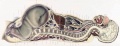
 Bailey178.jpg 913 × 653; 125 KB
 Bailey334.jpg 857 × 558; 77 KB
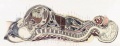
 Bailey447.jpg 795 × 423; 45 KB
 Bailey499.jpg 805 × 477; 65 KB
 Bailey500.jpg 1,215 × 677; 159 KB
 Bailey501.jpg 688 × 1,093; 276 KB
 Bailey502.jpg 751 × 547; 74 KB
 Braune 1877 plate 29B.jpg 868 × 1,200; 322 KB
 Braune 1877 plate 30.jpg 857 × 1,200; 295 KB
 Braune 1877 plate 31.jpg 870 × 1,200; 299 KB
 Braune2 C1.jpg 3,123 × 1,200; 918 KB
 BrauneA.jpg 1,200 × 485; 141 KB
 BrauneA1.jpg 1,200 × 485; 141 KB
 BrauneB1.jpg 1,200 × 485; 139 KB
 BrauneB2.jpg 1,200 × 485; 143 KB
 BrauneC1.jpg 1,200 × 461; 143 KB
 BrauneC2.jpg 1,200 × 461; 142 KB
 BrauneC3.jpg 1,200 × 461; 143 KB
 Dev anat 01.jpg 500 × 375; 25 KB
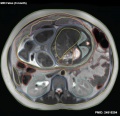
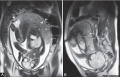
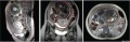 Fetal 9 month MRI 01.jpg 1,501 × 483; 145 KB
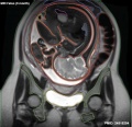
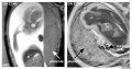
 Fetal 9 month MRI 02.jpg 619 × 600; 67 KB
 Fetal 9 month MRI 03.jpg 619 × 600; 64 KB
 Fetal 9 month MRI 04.jpg 619 × 600; 54 KB
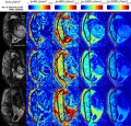
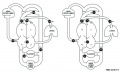 Fetal blood flow 01.jpg 1,000 × 599; 75 KB
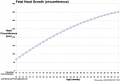 Fetal head growth circumference graph01.jpg 905 × 613; 58 KB
 Fetal length change.jpg 972 × 648; 72 KB
 Frazer006 bw600.jpg 600 × 575; 47 KB
 Human fetal adrenal GA32 large.mp4 ; 2.62 MB
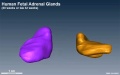 Human fetal adrenal GA32.jpg 800 × 502; 39 KB
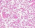
 Human fetal adrenal gland 01.jpg 1,266 × 800; 107 KB
 Hypospadia 3D ultrasound 01.jpg 1,150 × 497; 87 KB
 Keibel Mall 2 621.jpg 1,280 × 1,842; 589 KB
 Keith1902 fig094.jpg 800 × 646; 102 KB
 Kollmann618.jpg 940 × 592; 50 KB
 Kollmann619.jpg 750 × 639; 51 KB
 Kollmann620.jpg 772 × 613; 50 KB
 Kollmann621.jpg 976 × 550; 49 KB
 Lockwood1888b fig49.jpg 800 × 496; 69 KB
 Placenta MRI 01.jpg 800 × 516; 60 KB
 Placenta MRI 02.jpg 1,200 × 631; 91 KB
 Placenta MRI01.jpg 1,280 × 1,227; 317 KB
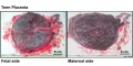 Placenta term anatomy 01.jpg 1,200 × 600; 115 KB
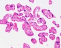 Placental villi 3.jpg 1,280 × 1,024; 99 KB
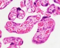 Placental villi 4.jpg 1,280 × 1,024; 89 KB
 Placental villi 5.jpg 1,280 × 1,024; 238 KB
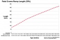
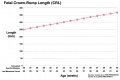 Third trimester Crown-Rump Length graph.jpg 972 × 648; 59 KB


