Sensory - Touch Development: Difference between revisions
mNo edit summary |
mNo edit summary |
||
| Line 170: | Line 170: | ||
{{#pmid:27702783}} | {{#pmid:27702783}} | ||
{{#pmid:27457037}} | |||
{{#pmid:25480024}} | {{#pmid:25480024}} | ||
Latest revision as of 10:47, 19 November 2019
| Embryology - 8 Jun 2024 |
|---|
| Google Translate - select your language from the list shown below (this will open a new external page) |
|
العربية | català | 中文 | 中國傳統的 | français | Deutsche | עִברִית | हिंदी | bahasa Indonesia | italiano | 日本語 | 한국어 | မြန်မာ | Pilipino | Polskie | português | ਪੰਜਾਬੀ ਦੇ | Română | русский | Español | Swahili | Svensk | ไทย | Türkçe | اردو | ייִדיש | Tiếng Việt These external translations are automated and may not be accurate. (More? About Translations) |
Introduction

These notes introduce the development of the sense of touch, part of the somatosensory system. Because of the distribution of the sensory structures within the skin, this topic is generally also covered in integumentary development and neural component is covered in peripheral nervous system development from neural crest.
| Touch: touch receptors | touch pathway | pacinian corpuscle | Meissner's corpuscle | Merkel cell | sensory modalities | neural crest | neural | Student project | integumentary |
| Senses Links: Introduction | placode | Hearing and Balance hearing | balance | vision | smell | taste | touch | Stage 22 | Category:Sensory |
Some Recent Findings
|
| More recent papers |
|---|
 This table allows an automated computer search of the external PubMed database using the listed "Search term" text link.
More? References | Discussion Page | Journal Searches | 2019 References | 2020 References Search term: Touch Development <pubmed limit=5>Touch Development</pubmed> |
| Older papers |
|---|
| These papers originally appeared in the Some Recent Findings table, but as that list grew in length have now been shuffled down to this collapsible table.
See also the Discussion Page for other references listed by year and References on this current page.
|
Historic People

- Georg Meissner - (1829-1905) German histologist, physiologist and anatomist. Beiträge zur Anatomie und Physiologie der Haut. (Contributions to the anatomy and physiology of the skin.) Leipzig, 1853.
- Friedrich Sigmund Merkel- (1845-1919) German anatomist and histologist, the name "Merckel cell" was based upon his first full description of touch cells (Tastzellen) and named by Robert Bonnet (1851–1921). Referred to these cells as Tastzellen or "touch cells" but this proposed function has been controversial as it has been hard to prove.
- Filippo Pacini - (1812-1883) Italian anatomist, published in 1840, and the name "Pacini's corpuscles" was proposed in 1844 by Henle and also by Kölliker.
- Angelo Ruffini (1864-1929) Italian histologist and embryologist, the name "Ruffini corpuscles" was based upon his first description of skin mechanoreceptors.
Touch Receptors


Touch receptors in mammalian skin and the neural encoding of reception.[1]
Touch Pathway
Pacinian Corpuscle
These receptors are lamellar vibration receptors that produce rapidly adapting responses. They are located in the subcutaneous tissue, deeper in interosseous membranes, and also mesenteries of the gut. The inner core cells form from Schwann cells extending from the nerve terminal.
A recent study in developing human digits,[3] show appearance at 11 weeks (13 weeks GA), and completed at 4 months of life. Both basic structure and immunohistochemical characteristics were reached at 36 weeks GA. The Schwann cell processes progressively compact to form the inner core, while the surrounding mesenchyme is organised to form the outer core component and the capsule.

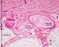
Pacinian corpuscle (labeled)
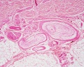
Pacinian corpuscle (unlabeled)
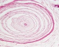
Pacinian corpuscle (detail)


Meissner's Corpuscle
Meissner's corpuscles are mechanoreceptors located in glabrous skin within the dermal papillae for sensitivity to light touch. They are rapidly adaptive elongated receptors consisting of a connective tissue capsule containing several lamellae of Schwann cells enclosing one or more afferent nerve fibres. The sensory corpuscle is a stack of discoid components of flattened axon terminals sandwiched between Schwann cell lamellae covered with a connective tissue capsule linked to the basal aspect of the epidermis by dermal collagen fibers.[12]
A recent study in developing human digits,[3] show appearance at 20 weeks (22 weeks GA and complete their typical morphology and immunohistochemical profile at 8 months of life.


Meissner's corpuscles are located throughout the skin and are seen concentrated in regions of high touch (finger tips, lips, genital regions). There is evidence that these receptors are concentrated in the fingertips of primates and may be associated with the evolution of manipulative abilities of the hands.[13]
Structure
Coiled spring-like structures, composed of stacked, disk-like lamellar cells with lamellae orientation usually parallel to the skin surface.
- lamellar cells - Schwann cell-derived with peripherally displaced nuclei in a fibroblastic capsule incomplete at the apex.
- sensory neurites - course through the lamellae, not visible by (Stain - Haematoxylin Eosin).
Merkel Cell
Merkel cells develop from epidermal progenitor population differentiation in the embryo, and also during their replacement postnatally.[14] They form a long-lived cell population within the skin with both a sensory and a neuroendocrine functions.
Merkel cells occur in many species including reptiles, fish, and mammals. Merkel cell complexes mediate slowly adapting type I (SAI) responses, which are characterized by an irregular firing pattern during sustained pressure. Merkel cells arise in the embryo[8] and in the adult[7] from an epidermal progenitor cell population.[15] These cells express the transcription factor Atoh1 and when differentiated are post-mitotic. Merkel cells can be organised into specialised clustered structure known as a "touch dome" associated with primary hair follicles.

A molecular signalling cascade from Wnt, Eda, and Shh is required for touch dome Merkel cell development.[6] Keratin 8 (K8) and Keratin 18 (K18) are Merkel cell markers identified in late fetal and adult skin.
The Notch signaling pathway has been shown to antagonize Atoh1 expression that is required for Merkel cell development in the skin. [2]

Isolated Merkel cell (em) |
Merkel cell (Merkel-Ranvier cell) integumentary (skin) receptor cell connected with somatosensory afferents.
Cell characteristics a polylobulated nucleus and numerous typical dense-core granules in a clear cytoplasm. The name "Merckel cell" was based upon Friedrich Sigmund Merkel (1845 – 1919) a German anatomist and histologist, first description of these touch cells (Tastzellen) and named in his honour by Robert Bonnet (1851–1921). Scale bar 5 µm (Stain - Osmium) |

Integumentary touch dome suggested functions[16] |
The Merkel cell is also a part of the touch dome (TD) apparatus, an innervated structure composed of specialised keratinocytes, and may have additional neuroendocrine and immune roles, as they associate with Langerhans cells (dendritic antigen presenting cells) in the epidermis.[16] |
In the adult, abnormalities in Merkel cell development can lead to the rare disease of Merkel cell carcinoma (MCC) associated with sun (UV) exposure.
Sensory Modalities
Thermoreceptors
- Receptors for heat (warmth) and cold (chill) detection.
- heat - C-fibres
- cold - Aδ fibres
Nociceptors
- Receptors for encoding and processing noxious stimuli.
- A-δ nociceptors
- Polymodal C-nociceptors
- C- mechano-insensitive nociceptors
Abnormalities
- Merkel cell carcinoma (MCC) PMID 12007193
Historic
The fine structure of Meissner's touch corpuscles of human fingers.[17]
- "The principal part of Meissner's corpuscle is made up of flattened laminar cells stretching across the corpuscle in irregular layers. The perinuclear cytoplasm of these cells contains numerous small mitochondria, a sparse granular endoplasmic reticulum, and a large number of small vesicles. Nerve fibers enter the side or base of the corpuscle, lose their myelin sheaths, and follow a meandering course between the laminar cell plates. The nerve endings enter into a close appositional relationship with the flattened portions of the laminar cells. In some areas the apposed axolemma and cell membranes are slightly thickened with small vesicles located along the cell membrane or on both surfaces. These regions are interpreted as synapses. The most prominent feature of the nerve endings is an extraordinary accumulation of small mitochondria which vary in size and internal density. The nerve endings also contain vacuoles, groups of dense concentric membranes, and small dense vesicles of irregular distribution. The laminar cells are separated from one another by a dense intercellular substance of uniform thickness which also envelops the entire corpuscle. This material contains randomly oriented collagen fibers and fine fibrils bound together by a dense material at nodal points recurring at regular intervals of approximately 120 mmicro. These findings are discussed in relation to the problems of the function of Meissner's corpuscle, neural material loss and replacement, and the presence of synapses."
References
- ↑ 1.0 1.1 Lumpkin EA, Marshall KL & Nelson AM. (2010). The cell biology of touch. J. Cell Biol. , 191, 237-48. PMID: 20956378 DOI.
- ↑ 2.0 2.1 Logan GJ, Wright MC, Kubicki AC & Maricich SM. (2018). Notch pathway signaling in the skin antagonizes Merkel cell development. Dev. Biol. , 434, 207-214. PMID: 29241683 DOI.
- ↑ 3.0 3.1 3.2 Feito J, García-Suárez O, García-Piqueras J, García-Mesa Y, Pérez-Sánchez A, Suazo I, Cabo R, Suárez-Quintanilla J, Cobo J & Vega JA. (2018). The development of human digital Meissner's and Pacinian corpuscles. Ann. Anat. , 219, 8-24. PMID: 29842990 DOI.
- ↑ Kobayashi K, Cho KH, Yamamoto M, Mitomo K, Murakami G, Abe H & Abe S. (2018). Tree of Vater-Pacinian corpuscles in the human finger and thumb: a comparison between the late fetal stage and old age. Surg Radiol Anat , 40, 243-257. PMID: 28653179 DOI.
- ↑ Chen YC, Lewis TL, Shore DI, Spence C & Maurer D. (2018). Developmental changes in the perception of visuotactile simultaneity. J Exp Child Psychol , 173, 304-317. PMID: 29783043 DOI.
- ↑ 6.0 6.1 Xiao Y, Thoresen DT, Miao L, Williams JS, Wang C, Atit RP, Wong SY & Brownell I. (2016). A Cascade of Wnt, Eda, and Shh Signaling Is Essential for Touch Dome Merkel Cell Development. PLoS Genet. , 12, e1006150. PMID: 27414798 DOI.
- ↑ 7.0 7.1 Wright MC, Reed-Geaghan EG, Bolock AM, Fujiyama T, Hoshino M & Maricich SM. (2015). Unipotent, Atoh1+ progenitors maintain the Merkel cell population in embryonic and adult mice. J. Cell Biol. , 208, 367-79. PMID: 25624394 DOI.
- ↑ 8.0 8.1 Perdigoto CN, Bardot ES, Valdes VJ, Santoriello FJ & Ezhkova E. (2014). Embryonic maturation of epidermal Merkel cells is controlled by a redundant transcription factor network. Development , 141, 4690-6. PMID: 25468937 DOI.
- ↑ Ackerley R, Olausson H, Wessberg J & McGlone F. (2012). Wetness perception across body sites. Neurosci. Lett. , 522, 73-7. PMID: 22710006 DOI.
- ↑ Fabrizi L, Slater R, Worley A, Meek J, Boyd S, Olhede S & Fitzgerald M. (2011). A shift in sensory processing that enables the developing human brain to discriminate touch from pain. Curr. Biol. , 21, 1552-8. PMID: 21906948 DOI.
- ↑ Woo SH, Stumpfova M, Jensen UB, Lumpkin EA & Owens DM. (2010). Identification of epidermal progenitors for the Merkel cell lineage. Development , 137, 3965-71. PMID: 21041368 DOI.
- ↑ Takahashi-Iwanaga H & Shimoda H. (2003). The three-dimensional microanatomy of Meissner corpuscles in monkey palmar skin. J. Neurocytol. , 32, 363-71. PMID: 14724379 DOI.
- ↑ Verendeev A, Thomas C, McFarlin SC, Hopkins WD, Phillips KA & Sherwood CC. (2015). Comparative analysis of Meissner's corpuscles in the fingertips of primates. J. Anat. , 227, 72-80. PMID: 26053332 DOI.
- ↑ Van Keymeulen A, Mascre G, Youseff KK, Harel I, Michaux C, De Geest N, Szpalski C, Achouri Y, Bloch W, Hassan BA & Blanpain C. (2009). Epidermal progenitors give rise to Merkel cells during embryonic development and adult homeostasis. J. Cell Biol. , 187, 91-100. PMID: 19786578 DOI.
- ↑ Morrison KM, Miesegaes GR, Lumpkin EA & Maricich SM. (2009). Mammalian Merkel cells are descended from the epidermal lineage. Dev. Biol. , 336, 76-83. PMID: 19782676 DOI.
- ↑ 16.0 16.1 Xiao Y, Williams JS & Brownell I. (2014). Merkel cells and touch domes: more than mechanosensory functions?. Exp. Dermatol. , 23, 692-5. PMID: 24862916 DOI.
- ↑ CAUNA N & ROSS LL. (1960). The fine structure of Meissner's touch corpuscles of human fingers. J Biophys Biochem Cytol , 8, 467-82. PMID: 13691669
Reviews
Piccinin MA & Schwartz J. (2018). Histology, Meissner Corpuscle. , , . PMID: 30085522
Lai HC, Seal RP & Johnson JE. (2016). Making sense out of spinal cord somatosensory development. Development , 143, 3434-3448. PMID: 27702783 DOI.
Verriotis M, Chang P, Fitzgerald M & Fabrizi L. (2016). The development of the nociceptive brain. Neuroscience , 338, 207-219. PMID: 27457037 DOI.
Woo SH, Lumpkin EA & Patapoutian A. (2015). Merkel cells and neurons keep in touch. Trends Cell Biol. , 25, 74-81. PMID: 25480024 DOI.
Zimmerman A, Bai L & Ginty DD. (2014). The gentle touch receptors of mammalian skin. Science , 346, 950-4. PMID: 25414303 DOI.
Jeffry J, Kim S & Chen ZF. (2011). Itch signaling in the nervous system. Physiology (Bethesda) , 26, 286-92. PMID: 21841076 DOI.
Lumpkin EA, Marshall KL & Nelson AM. (2010). The cell biology of touch. J. Cell Biol. , 191, 237-48. PMID: 20956378 DOI.
CAUNA N & ROSS LL. (1960). The fine structure of Meissner's touch corpuscles of human fingers. J Biophys Biochem Cytol , 8, 467-82. PMID: 13691669
Articles
Ranade SS, Woo SH, Dubin AE, Moshourab RA, Wetzel C, Petrus M, Mathur J, Bégay V, Coste B, Mainquist J, Wilson AJ, Francisco AG, Reddy K, Qiu Z, Wood JN, Lewin GR & Patapoutian A. (2014). Piezo2 is the major transducer of mechanical forces for touch sensation in mice. Nature , 516, 121-5. PMID: 25471886 DOI.
Books
Neurobiology of Sensation and Reward. Gottfried JA, editor. Boca Raton (FL): CRC Press; 2011. Chapter 7 - Touch PMID 22593916
Search PubMed
Search Pubmed: Touch Development | touch receptors |
External Links
External Links Notice - The dynamic nature of the internet may mean that some of these listed links may no longer function. If the link no longer works search the web with the link text or name. Links to any external commercial sites are provided for information purposes only and should never be considered an endorsement. UNSW Embryology is provided as an educational resource with no clinical information or commercial affiliation.
- Merkel cell carcinoma - http://www.merkelcell.org
Terms
| Touch Terms | ||
|---|---|---|
|
Glossary Links
- Glossary: A | B | C | D | E | F | G | H | I | J | K | L | M | N | O | P | Q | R | S | T | U | V | W | X | Y | Z | Numbers | Symbols | Term Link
Cite this page: Hill, M.A. (2024, June 8) Embryology Sensory - Touch Development. Retrieved from https://embryology.med.unsw.edu.au/embryology/index.php/Sensory_-_Touch_Development
- © Dr Mark Hill 2024, UNSW Embryology ISBN: 978 0 7334 2609 4 - UNSW CRICOS Provider Code No. 00098G