Integumentary System - Gland Development: Difference between revisions
mNo edit summary |
mNo edit summary |
||
| Line 18: | Line 18: | ||
|-bgcolor="F5FAFF" | |-bgcolor="F5FAFF" | ||
| | | | ||
* '''Molecular dynamics of Dkk4 modulates Wnt action and regulates meibomian gland development'''{{#pmid:27864382|PMID27864382}} "Secreted Dickkopf (Dkk) proteins are major {{Wnt}} pathway modulators during organ development. Dkk1 has been widely studied and acts as a general Wnt inhibitor. However, the molecular function of other Dkks remains largely unknown. Here, we show that Dkk4 selectively inhibits a subset of Wnts, but is further inactivated by proteolytic cleavage. Meibomian gland (MG) formation is employed as a model where Dkk4 and its Wnt targets are expressed. Skin-specific expression of Dkk4 arrests MG growth at early germ phase, which is similar to that observed in Eda-ablated Tabby mice. Consistent with transient Dkk4 action, intact Dkk4 inhibits MG extension but the cleaved form progressively increases during MG development with a concomitant upswing in Wnt activity. Furthermore, both Dkk4 and its receptor (and Wnt co-receptor) Lrp6 are direct Eda targets during MG induction. In cell and organotypic cultures, Dkk4 inhibition is eliminated by elevation of Lrp6. Also, Lrp6 upregulation restores MG formation in Tabby mice. Thus, the dynamic state of Dkk4 itself and its interaction with Lrp6 modulates Wnt function during MG development, with a novel limitation of Dkk4 action by proteolytic cleavage." | |||
* '''Involvement of Wnt, Eda and Shh at defined stages of sweat gland development'''{{#pmid:25249463|PMID25249463}} "Sweat gland induction failed completely when canonical Wnt signaling was blocked in skin epithelium, and was accompanied by sharp downregulation of downstream Wnt, Eda and Shh pathway genes. The Wnt antagonist Dkk4 appeared to inhibit this induction: Dkk4 was sharply downregulated in β-catenin-ablated mice, indicating that it is induced by Wnt/β-catenin; however, its overexpression repressed Wnt target genes and significantly reduced gland numbers. Eda signaling succeeded Wnt. Wnt signaling was still active and nascent sweat gland pre-germs were still seen in Eda-null mice, but the pre-germs failed to develop further and the downstream Shh pathway was not activated. When Wnt and Eda were intact but Shh was ablated, germ induction and subsequent duct formation occurred normally, but the final stage of secretory coil formation failed. Thus, sweat gland development shows a relay of regulatory steps initiated by Wnt/β-catenin - itself modulated by Dkk4 - with subsequent participation of Eda and Shh pathways." (this same singling pathway is required for sensory Merkel cell development {{#pmid:27414798|PMID27414798}} ) | * '''Involvement of Wnt, Eda and Shh at defined stages of sweat gland development'''{{#pmid:25249463|PMID25249463}} "Sweat gland induction failed completely when canonical Wnt signaling was blocked in skin epithelium, and was accompanied by sharp downregulation of downstream Wnt, Eda and Shh pathway genes. The Wnt antagonist Dkk4 appeared to inhibit this induction: Dkk4 was sharply downregulated in β-catenin-ablated mice, indicating that it is induced by Wnt/β-catenin; however, its overexpression repressed Wnt target genes and significantly reduced gland numbers. Eda signaling succeeded Wnt. Wnt signaling was still active and nascent sweat gland pre-germs were still seen in Eda-null mice, but the pre-germs failed to develop further and the downstream Shh pathway was not activated. When Wnt and Eda were intact but Shh was ablated, germ induction and subsequent duct formation occurred normally, but the final stage of secretory coil formation failed. Thus, sweat gland development shows a relay of regulatory steps initiated by Wnt/β-catenin - itself modulated by Dkk4 - with subsequent participation of Eda and Shh pathways." (this same singling pathway is required for sensory Merkel cell development {{#pmid:27414798|PMID27414798}} ) | ||
Latest revision as of 14:14, 10 November 2019
| Embryology - 23 May 2024 |
|---|
| Google Translate - select your language from the list shown below (this will open a new external page) |
|
العربية | català | 中文 | 中國傳統的 | français | Deutsche | עִברִית | हिंदी | bahasa Indonesia | italiano | 日本語 | 한국어 | မြန်မာ | Pilipino | Polskie | português | ਪੰਜਾਬੀ ਦੇ | Română | русский | Español | Swahili | Svensk | ไทย | Türkçe | اردو | ייִדיש | Tiếng Việt These external translations are automated and may not be accurate. (More? About Translations) |
Introduction
| The secretory glands associated with the integumentary system have similar embryonic origins and mechanisms of development, though are specialised by their locations and secretions. A key process and feature of all gland development is an epithelial-mesenchymal interaction(s). Gland secretion can also be regulated by a number of different mechanisms, including endocrine changes postnatally at puberty and during pregnancy.
|

|

|

|
Some Recent Findings
|
| More recent papers |
|---|
 This table allows an automated computer search of the external PubMed database using the listed "Search term" text link.
More? References | Discussion Page | Journal Searches | 2019 References | 2020 References Search term: Gland Embryology | Integumentary Gland Development |
Textbooks
- Human Embryology (2nd ed.) Larson Chapter 14 p443-455
- The Developing Human: Clinically Oriented Embryology (6th ed.) Moore and Persaud Chapter 20: P513-529
- Before We Are Born (5th ed.) Moore and Persaud Chapter 21: P481-496
- Essentials of Human Embryology Larson Chapter 14: P303-315
- Human Embryology, Fitzgerald and Fitzgerald
- Color Atlas of Clinical Embryology Moore Persaud and Shiota Chapter 15: p231-236
Development of Glands
Two main surface types, sweat and sebaceous, with a number of specialized glands in integumentary regions (lacrimal, Meibomian).
- both ectodermal in origin
- form as ingrowth of ectoderm into the mesoderm
Secretion Mechanisms
| Merocrine | Apocrine | Holocrine |
|---|---|---|
|
|
|
|
Sweat Gland
Humans have two types of sweat glands are present in humans, differing in secretory mechanism, number, histological appearance and sweat composition.
- merocrine (~eccrine) sweat glands (majority)
- apocrine sweat glands (minority)
- apocrine in axilla, pubic and nipple regions
- see also mammary gland development
Skin merocrine sweat gland
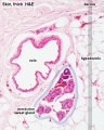
Skin merocrine sweat gland
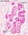
Skin merocrine sweat gland
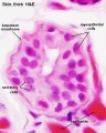
Skin merocrine sweat gland (detail)


Sebaceous Gland


Adult skin sebaceous gland histology
- associated with hair development
- except plans penis and labia minora
- these glands secrete vernix
Vernix Caseosa

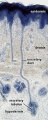
(vernix, Latin, vernix = varnish, caseous = cheese=like) This is a specialized coating that forms in late development over the entire fetal surface. The main component, secreted sebum, is secreted by sebaceous glands. The other constituents are cells sloughed off the fetus's skin, and shed lanugo hair. The coating also has a high water content (80%) largely compartmentalized within fetal corneocytes (cells forming the stratum corneum).[5]
This coating develops intially in a cranio-caudal direction and can be absent in preterm infants.
Some functions include:
- protection of the fetal skin from extraembryonic fluids amnion, urine
- providing a slippery surface helps with parturition (birth)
- acting as a biofilm barrier against infection
Lacrimal Glands
Lacrimal glands produce the aqueous tears that mix with Meibomian glands secretions to form a tear film coating the outer surface of the eye.
Human Development (based upon data from[6])
- Carnegie stage 16 - 18 - formation of the lacrimal lamina
- Carnegie stage 19 - 23 - formation of the lacrimal cord
- fetal Week 9+ - maturation of the excretory lacrimal system
- Links: vision
Meibomian Glands
The Meibomian glands (glandulae tarsales) are sebaceous glands located at the margins of the upper and lower eyelids of humans and mammals.[7] The gland cells (meibocytes) secrete by a holocrine mechanism and produce constantly a lipid-rich secretion (meibum) that mix with with aqueous tears produced by lacrimal glands. These glands are also regulated through sex hormones, androgens have a supporting function while estrogens act antagonistically.
The glands were first described in 1666 by Heinrich Meibom (1638 - 1700) a German physician and anatomist.
Abnormalities
Alacrima
Term describes a range of abnormalities associated with lacrimal gland development. The most common genetic cause of alacrima is the Riley-Day syndrome.
Nasolacrimal Duct Obstruction
Drainage duct obstruction can be a common anomaly in children and can be caused by inflammation or fibrosis without any precipitating cause (primary classification) or by an acquired lacrimal drainage obstruction (secondary classification).
Vernix Caseosa Peritonitis
Not an abnormality of development, but a clinical term for a rare post-caesarean section complication due to spilt vernix caseosa at the time of caesarean section mediating a maternal inflammatory reaction.[8]
References
- ↑ Sima J, Piao Y, Chen Y & Schlessinger D. (2016). Molecular dynamics of Dkk4 modulates Wnt action and regulates meibomian gland development. Development , 143, 4723-4735. PMID: 27864382 DOI.
- ↑ Cui CY, Yin M, Sima J, Childress V, Michel M, Piao Y & Schlessinger D. (2014). Involvement of Wnt, Eda and Shh at defined stages of sweat gland development. Development , 141, 3752-60. PMID: 25249463 DOI.
- ↑ Xiao Y, Thoresen DT, Miao L, Williams JS, Wang C, Atit RP, Wong SY & Brownell I. (2016). A Cascade of Wnt, Eda, and Shh Signaling Is Essential for Touch Dome Merkel Cell Development. PLoS Genet. , 12, e1006150. PMID: 27414798 DOI.
- ↑ Chen WC & Zouboulis CC. (2009). Hormones and the pilosebaceous unit. Dermatoendocrinol , 1, 81-6. PMID: 20224689
- ↑ Pickens WL, Warner RR, Boissy YL, Boissy RE & Hoath SB. (2000). Characterization of vernix caseosa: water content, morphology, and elemental analysis. J. Invest. Dermatol. , 115, 875-81. PMID: 11069626 DOI.
- ↑ de la Cuadra-Blanco C, Peces-Peña MD, Jáñez-Escalada L & Mérida-Velasco JR. (2006). Morphogenesis of the human excretory lacrimal system. J. Anat. , 209, 127-35. PMID: 16879594 DOI.
- ↑ Butovich IA. (2009). The Meibomian puzzle: combining pieces together. Prog Retin Eye Res , 28, 483-98. PMID: 19660571 DOI.
- ↑ Stuart OA, Morris AR & Baber RJ. (2009). Vernix caseosa peritonitis - no longer rare or innocent: a case series. J Med Case Rep , 3, 60. PMID: 19208257 DOI.
Journals
Reviews
Takahashi Y, Kakizaki H, Chan WO & Selva D. (2010). Management of congenital nasolacrimal duct obstruction. Acta Ophthalmol , 88, 506-13. PMID: 19681790 DOI.
Weber AL, Rodriguez-DeVelasquez A, Lucarelli MJ & Cheng HM. (1996). Normal anatomy and lesions of the lacrimal sac and duct: evaluated by dacryocystography, computed tomography, and MR imaging. Neuroimaging Clin. N. Am. , 6, 199-217. PMID: 8919141
Moore BD. (1994). Lacrimal system abnormalities. Optom Vis Sci , 71, 182-3. PMID: 8196943
Rowzee AM, Lazzarino DA, Rota L, Sun Z & Wood TL. (2008). IGF ligand and receptor regulation of mammary development. J Mammary Gland Biol Neoplasia , 13, 361-70. PMID: 19020961 DOI.
Articles
Singh G & Archana G. (2008). Unraveling the mystery of vernix caseosa. Indian J Dermatol , 53, 54-60. PMID: 19881987 DOI.
Tollin M, Bergsson G, Kai-Larsen Y, Lengqvist J, Sjövall J, Griffiths W, Skúladóttir GV, Haraldsson A, Jörnvall H, Gudmundsson GH & Agerberth B. (2005). Vernix caseosa as a multi-component defence system based on polypeptides, lipids and their interactions. Cell. Mol. Life Sci. , 62, 2390-9. PMID: 16179970 DOI.
Search PubMed
Search Pubmed: Epithelial Gland Development | Sweat Gland Development | Sebaceous Gland Development | Eccrine Gland Development | Apocrine Gland Development | Lacrimal Gland Development | Meibomian Gland Development
Additional Images
Terms
| Integumentary Terms | ||
|---|---|---|
Integumentary Development
| ||
|
External Links
External Links Notice - The dynamic nature of the internet may mean that some of these listed links may no longer function. If the link no longer works search the web with the link text or name. Links to any external commercial sites are provided for information purposes only and should never be considered an endorsement. UNSW Embryology is provided as an educational resource with no clinical information or commercial affiliation.
Glossary Links
- Glossary: A | B | C | D | E | F | G | H | I | J | K | L | M | N | O | P | Q | R | S | T | U | V | W | X | Y | Z | Numbers | Symbols | Term Link
Cite this page: Hill, M.A. (2024, May 23) Embryology Integumentary System - Gland Development. Retrieved from https://embryology.med.unsw.edu.au/embryology/index.php/Integumentary_System_-_Gland_Development
- © Dr Mark Hill 2024, UNSW Embryology ISBN: 978 0 7334 2609 4 - UNSW CRICOS Provider Code No. 00098G