File:Hertig1956 fig20.jpg: Difference between revisions
No edit summary |
mNo edit summary |
||
| Line 1: | Line 1: | ||
==Fig. 20.== | |||
Three 9-day specimens of Horizon Vb, characterized by syncytiotrophoblastic lacunae with early utero-placental circulation, amniogenesis, a simple bilaminar germ disc and only variable degrees of exocoelomie (Heuser’s) membrane formation. | |||
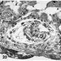
20 Detail of embryo and surrounding trophoblast. Note bilaminar germ disc with vacuolated ectoderm, early amniogenesis and an early exoeoelomic membrane. delaminating from the cytotrophoblast, best seen at the abembryonic pole. Note small shallow surface ulcer with alteration of adjacent nuclei. The paler cytotrophoblast lines the chorionie cavity and varies in thickness whereas the darker synrcytiotrophoblast lies peripherally and contains many intracytoplasmic lacunae which are coalescing to form the future intcrvillous space. Note mitotic figure in cytotrophoblast. [[:Category:Carnegie Embryo 8171|Carnegie 8171]], Section 3-:2-11. X 300. | |||
{{Hertig1956 figures}} | |||
Latest revision as of 20:25, 23 February 2017
Fig. 20.
Three 9-day specimens of Horizon Vb, characterized by syncytiotrophoblastic lacunae with early utero-placental circulation, amniogenesis, a simple bilaminar germ disc and only variable degrees of exocoelomie (Heuser’s) membrane formation.
20 Detail of embryo and surrounding trophoblast. Note bilaminar germ disc with vacuolated ectoderm, early amniogenesis and an early exoeoelomic membrane. delaminating from the cytotrophoblast, best seen at the abembryonic pole. Note small shallow surface ulcer with alteration of adjacent nuclei. The paler cytotrophoblast lines the chorionie cavity and varies in thickness whereas the darker synrcytiotrophoblast lies peripherally and contains many intracytoplasmic lacunae which are coalescing to form the future intcrvillous space. Note mitotic figure in cytotrophoblast. Carnegie 8171, Section 3-:2-11. X 300.
- Figure Links: 1 | 2 | 3 | 4 | 5 | 6 | 7 | 8 | 9-10 | 11-12 | 13-14 | 15-16 | 17 | 18-19 | 20 | 21-22 | 23 | 24-25 | 26-27 | 28-29 | 30-31 | 32-33 | 34 | 35 | 36 | 37 | 38 | 39 | 40 | 41 | 42 | 43 | 44 | 45 | 46 | 47 | 48 | 40 | 49 | 50 | 51 | 52 | 53 | 54 | 55 | 56 | 57 | 58 | 59 | 60 | 61 | 62 | 63 | 64 | 65 | 66 | 67 | 68 | 69 | 70 | 71 | 72 | 73 | 74 | 75 | 76 | 77 | 78 | 79 | 80 | 81 | 82 | 83 | 84 | 85 | 86 | 87 | 88 | 89 | 90 | plate 1 | plate 2 | plate 3 | plate 4 | plate 5 | plate 6 | plate 7 | plate 8 | plate 9 | plate 10 | plate 11 | plate 12 | plate 13 | plate 14 | plate 15 | plate 16 | plate 17 | table 1 | table 1 image | table 2 image | table 3 image | table 4 | table 4 image | table 5 | table 5 image | All figures | 1956 Hertig | Embryology History - Arthur Hertig | John Rock | Historic Papers
Reference
Hertig AT. Rock J. and Adams EC. A description of 34 human ova within the first 17 days of development. (1956) Amer. J Anat., 98:435-493.
Cite this page: Hill, M.A. (2024, June 6) Embryology Hertig1956 fig20.jpg. Retrieved from https://embryology.med.unsw.edu.au/embryology/index.php/File:Hertig1956_fig20.jpg
- © Dr Mark Hill 2024, UNSW Embryology ISBN: 978 0 7334 2609 4 - UNSW CRICOS Provider Code No. 00098G
File history
Click on a date/time to view the file as it appeared at that time.
| Date/Time | Thumbnail | Dimensions | User | Comment | |
|---|---|---|---|---|---|
| current | 20:22, 23 February 2017 |  | 700 × 696 (110 KB) | Z8600021 (talk | contribs) |
You cannot overwrite this file.
File usage
The following 3 pages use this file: