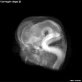
Carnegie stage 23: Difference between revisions
m (→Events) |
m (→Events) |
||
| Line 81: | Line 81: | ||
* Renal - Metanephros secretory tubules are changing from short to long, and becoming more convoluted. The epithelium in some tubules is high. Renal tubules of fourth and fifth orders are present. Large glomeruli are numerous (fig. 19-12). | * Renal - Metanephros secretory tubules are changing from short to long, and becoming more convoluted. The epithelium in some tubules is high. Renal tubules of fourth and fifth orders are present. Large glomeruli are numerous (fig. 19-12). | ||
* Humerus. All five cartilaginous phases are now present (Streeter, 1949, figs. 3 and 18). | * Humerus. All five cartilaginous phases are now present (Streeter, 1949, figs. 3 and 18). | ||
* [[Endocrine System Development]]<ref name=O'Rahilly1983a>{{Ref-O'Rahilly1983a}}</ref> | * [[Endocrine System Development|'''Endocrine''']]<ref name=O'Rahilly1983a>{{Ref-O'Rahilly1983a}}</ref> | ||
** Pituitary - adenohypophysis loss of the stalk and lobules of epithelium project into the mesodermal component of the gland, and oriented epithelial follicles are present (Streeter, 1951, plate 2). Abundant angioblasts and capillaries are found. | ** Pituitary - adenohypophysis loss of the stalk and lobules of epithelium project into the mesodermal component of the gland, and oriented epithelial follicles are present (Streeter, 1951, plate 2). Abundant angioblasts and capillaries are found. | ||
** Epiphysis - The pineal body has reached Stadium 5 of Turkewitsch (1933) (O'Rahilly 1968). <ref name=O'Rahilly1968>{{Ref-O'Rahilly1968}}</ref> | ** Epiphysis - The pineal body has reached Stadium 5 of Turkewitsch (1933) (O'Rahilly 1968). <ref name=O'Rahilly1968>{{Ref-O'Rahilly1968}}</ref> | ||
Revision as of 17:37, 31 October 2016
| Embryology - 14 Jun 2024 |
|---|
| Google Translate - select your language from the list shown below (this will open a new external page) |
|
العربية | català | 中文 | 中國傳統的 | français | Deutsche | עִברִית | हिंदी | bahasa Indonesia | italiano | 日本語 | 한국어 | မြန်မာ | Pilipino | Polskie | português | ਪੰਜਾਬੀ ਦੇ | Română | русский | Español | Swahili | Svensk | ไทย | Türkçe | اردو | ייִדיש | Tiếng Việt These external translations are automated and may not be accurate. (More? About Translations) |
Introduction

This is the final Carnegie stage of embryonic development in Week 8. After this development is considered fetal for the remainder of the pregnancy.
Facts
Week 8, 56 - 60 days, 27 - 31 mm
Gestational age GA week 10
Summary
- Ectoderm:
- Mesoderm: ossification continues
- Head: eyelids, external ears, rounded head
- Body: straightening of trunk, intestines herniated at umbilicus
- Limbs: hands and feet turned inward
See also Carnegie stage 23 Events
Features
- scalp vascular plexus, eylid, eye, nose, auricle of external ear, mouth, shoulder, arm, elbow, wrist, toes separated, sole of foot, umbilical cord
- hearing - cochlea shows nearly 21/2 turns. otic capsule cartilage separated from the semicircular ducts by a pre cartilaginous zone. Labyrinth has practically completed its gross development and ductus reuniens is well defined.[1]
- Links: Week 8 | System Development | Lecture - Limb | Lecture - Head Development | Lecture - Sensory | Science Practical - Head | Science Practical - Sensory | Science Practical - Urogenital | Category:Carnegie Stage 23 | Fetal Development
| Week: | 1 | 2 | 3 | 4 | 5 | 6 | 7 | 8 |
| Carnegie stage: | 1 2 3 4 | 5 6 | 7 8 9 | 10 11 12 13 | 14 15 | 16 17 | 18 19 | 20 21 22 23 |
- Carnegie Stages: 1 | 2 | 3 | 4 | 5 | 6 | 7 | 8 | 9 | 10 | 11 | 12 | 13 | 14 | 15 | 16 | 17 | 18 | 19 | 20 | 21 | 22 | 23 | About Stages | Timeline
Kyoto Collection

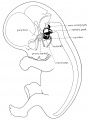
View: This is a dorsolateral view of embryo. Amniotic membrane removed.
Image source: Embryology page Created: 19.03.1999
Image source: The Kyoto Collection images are reproduced with the permission of Prof. Kohei Shiota and Prof. Shigehito Yamada, Anatomy and Developmental Biology, Kyoto University Graduate School of Medicine, Kyoto, Japan for educational purposes only and cannot be reproduced electronically or in writing without permission.
Carnegie Collection


- Carnegie stage 23: 4570 right | 4570 anterior | 4570 left | 4570 posterior | 4570 large right | 4570 large left | Rotation | Carnegie Embryos

| iBook - Carnegie Embryos | |
|---|---|

|
|
Hill Collection
| Hill HH12 | |
|---|---|

|

|

|

|
- Links: Hill Collection
Events
- Vision
- The retina comprises the pigmented layer, external limiting membrane, proliferative zone, external neuroblastic layer, transient fiber layer, internal neuroblastic layer, nerve fiber layer, and internal limiting membrane. Eyelids closure is complete (Note - shown as still open in the Kyoto embryo).[3]
- The cornea now comprises the anterior epithelium and its basement membrane, the substantia propria, and the posterior epithelium (Streeter, 1951, fig. 18, and O’Rahilly, 1966, figs. 51 and 59).
- Optic nerve a vascular canal is present and sheath in the more advanced specimens (O’Rahilly, 1966, fig. 55).
- Joint - knee cavity, anterior cruciate ligament and posterior cruciate ligament present[4]
- Hearing - Cochlear duct tip now points “downward” for the second time (fig. 19-6). The duct is coiled to nearly its final extent of 2½ turns.
- Vomeronasal organ. A narrow canal is seen in the long, tapering duct. The sac is beginning to shrink and retrogress (fig. 19-9).
- Submandibular gland. Lumina are found in many terminal branches of the duct (fig. 19-10). Orientation of the epithelial tree is beginning, and angiogenesis is commencing around the epithelium. A mesodermal sheath is beginning to form around the gland.
- Renal - Metanephros secretory tubules are changing from short to long, and becoming more convoluted. The epithelium in some tubules is high. Renal tubules of fourth and fifth orders are present. Large glomeruli are numerous (fig. 19-12).
- Humerus. All five cartilaginous phases are now present (Streeter, 1949, figs. 3 and 18).
- Endocrine[5]
- Pituitary - adenohypophysis loss of the stalk and lobules of epithelium project into the mesodermal component of the gland, and oriented epithelial follicles are present (Streeter, 1951, plate 2). Abundant angioblasts and capillaries are found.
- Epiphysis - The pineal body has reached Stadium 5 of Turkewitsch (1933) (O'Rahilly 1968). [6]
- Thymus - The cortex is well-developed, "true lobulation" has begun with the appearance of" fine superficial scallops," lymphocytes are present sparsely in the subcortical zone, and vessels are found within the thymus (Norris 1938).
- Adrenal Cortex - It appears that C2 cells first enter the body of the gland at this stage. The pattern of the arterial supply is established. The cellular "capsule" is penetrated by arterial capillaries which join the sinusoids. Their points of entry give the surface of the gland an appearance of cobblestones. The zona glomerulosa is formed of CI and C3 cells. Cells from this zone and from the "capsule" migrate centrally into the cords.[7]
- Adrenal Medulla - Nerve fibres and neuroblasts are first seen in the body of the gland. The paragangtion (M3) cells are beginning to multiply rapidly and, from 30 mm (stage 23) until birth, some are differentiating into chro- maffin cells.[7]
References
- ↑ Template:Ref-Streeter1906
- ↑ Streeter GL. Developmental Horizons In Human Embryos Description Or Age Groups XIX, XX, XXI, XXII, And XXIII, Being The Fifth Issue Of A Survey Of The Carnegie Collection. (1957) Carnegie Instn. Wash. Publ. 611, Contrib. Embryol., 36: 167-196.
- ↑ <pubmed>7364662</pubmed>
- ↑ <pubmed>9185992</pubmed>
- ↑ O'Rahilly R. The timing and sequence of events in the development of the human endocrine system during the embryonic period proper. (1983) Anat. Embryol., 166: 439-451. PMID 6869855
- ↑ O'Rahilly R. The development of the epiphysis cerebri and the subcommissural complex in staged human embryos. (1968) Anat. Rec., 160: 488-489.
- ↑ 7.0 7.1 Crowder RE. The development of the adrenal gland in man, with special reference to origin and ultimate location of cell types and evidence in favor of the "cell migration" theory. (1957) Contrib. Embryol., Carnegie Inst. Wash. 36, 193-210.
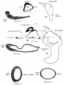
Additional Images
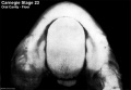
Oral cavity floor
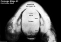
Oral cavity floor (labeled)

Oral cavity roof
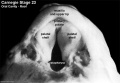
Oral cavity roof (labeled)

External ear Stages 14-23 and adult
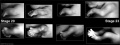
Stage 20-23 limbs
Stage 23 Optical Projection Tomography


Historic Images
| Historic Disclaimer - information about historic embryology pages |
|---|
 |
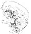
1906 Membranous labyrinth
1906 Semicircular canal
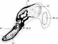
1908 Notochord
1910 Skull
1913 Clavicle


- Carnegie Stages: 1 | 2 | 3 | 4 | 5 | 6 | 7 | 8 | 9 | 10 | 11 | 12 | 13 | 14 | 15 | 16 | 17 | 18 | 19 | 20 | 21 | 22 | 23 | About Stages | Timeline
Cite this page: Hill, M.A. (2024, June 14) Embryology Carnegie stage 23. Retrieved from https://embryology.med.unsw.edu.au/embryology/index.php/Carnegie_stage_23
- © Dr Mark Hill 2024, UNSW Embryology ISBN: 978 0 7334 2609 4 - UNSW CRICOS Provider Code No. 00098G