File:Zebrafish brain fold SEM.jpg: Difference between revisions
(==Zebrafish Brain Fold SEM== Scanning EM of the zebrafish head folds. Specimens were chemically fixed critically point dried, and sputter coated with gold/palladium. This image is part of a series taken by [http://www.unb.ca/fredericton/science/biology) |
mNo edit summary |
||
| Line 6: | Line 6: | ||
Specimens were chemically fixed critically point dried, and sputter coated with gold/palladium. This image is part of a series taken by [http://www.unb.ca/fredericton/science/biology/Faculty/crawford/crawford.html Bryan Crawford] while he was at the University of Washington. They are part of the Zebrafish--The Living Laboratory CD made available by Mark Cooper and described in Methods in Cell Biology Volume 77, 2004, Pages 439-457. | Specimens were chemically fixed critically point dried, and sputter coated with gold/palladium. This image is part of a series taken by [http://www.unb.ca/fredericton/science/biology/Faculty/crawford/crawford.html Bryan Crawford] while he was at the University of Washington. They are part of the Zebrafish--The Living Laboratory CD made available by Mark Cooper and described in Methods in Cell Biology Volume 77, 2004, Pages 439-457. | ||
===Reference=== | |||
Mark Cooper '''Zebrafish - The Living Laboratory CD''' | |||
<pubmed>15602926</pubmed> | |||
====Copyright==== | |||
Licensing: Attribution Non-Commercial Share Alike:This image is licensed under a [http://creativecommons.org/licenses/by-nc-sa/3.0/ Creative Commons Attribution], Non-Commercial Share Alike License. | Licensing: Attribution Non-Commercial Share Alike:This image is licensed under a [http://creativecommons.org/licenses/by-nc-sa/3.0/ Creative Commons Attribution], Non-Commercial Share Alike License. | ||
Original image name:12660.jpg http://www.cellimagelibrary.org/images/12660 | |||
[[Category:Zebrafish]] [[Category:Scanning EM]] | [[Category:Zebrafish]] [[Category:Scanning EM]] | ||
Revision as of 09:24, 4 December 2014
Zebrafish Brain Fold SEM
Scanning EM of the zebrafish head folds.
Specimens were chemically fixed critically point dried, and sputter coated with gold/palladium. This image is part of a series taken by Bryan Crawford while he was at the University of Washington. They are part of the Zebrafish--The Living Laboratory CD made available by Mark Cooper and described in Methods in Cell Biology Volume 77, 2004, Pages 439-457.
Reference
Mark Cooper Zebrafish - The Living Laboratory CD
<pubmed>15602926</pubmed>
Copyright
Licensing: Attribution Non-Commercial Share Alike:This image is licensed under a Creative Commons Attribution, Non-Commercial Share Alike License.
Original image name:12660.jpg http://www.cellimagelibrary.org/images/12660
File history
Click on a date/time to view the file as it appeared at that time.
| Date/Time | Thumbnail | Dimensions | User | Comment | |
|---|---|---|---|---|---|
| current | 08:23, 26 April 2011 | 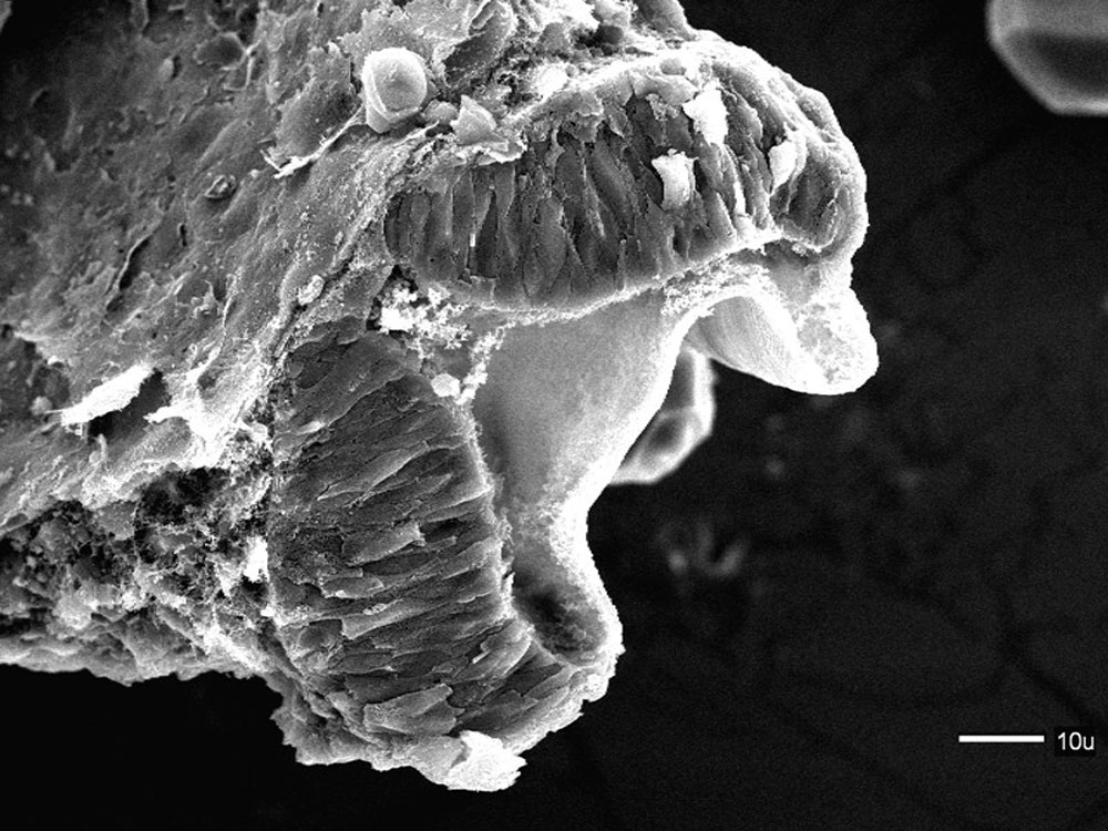 | 1,000 × 750 (172 KB) | S8600021 (talk | contribs) | ==Zebrafish Brain Fold SEM== Scanning EM of the zebrafish head folds. Specimens were chemically fixed critically point dried, and sputter coated with gold/palladium. This image is part of a series taken by [http://www.unb.ca/fredericton/science/biology |
You cannot overwrite this file.
File usage
The following page uses this file: