ANAT2241 Male Reproductive System: Difference between revisions
mNo edit summary |
|||
| Line 17: | Line 17: | ||
[http://vslides.unsw.edu.au/VirtualSlideV2.nsf/id/258A08 '''Virtual Slides''']: [http://vslides.unsw.edu.au/VirtualSlideV2.nsf/id/749C96 ANAT2241 Male Reproductive] | [http://vslides.unsw.edu.au/VirtualSlideV2.nsf/id/258A08 '''Virtual Slides''']: [http://vslides.unsw.edu.au/VirtualSlideV2.nsf/id/749C96 ANAT2241 Male Reproductive] | ||
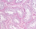
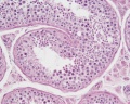
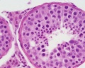
==Histology== | ==Testis Histology== | ||
{| | |||
| | |||
===Convoluted Seminiferous Tubules=== | |||
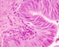
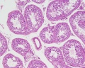
* tubules are enclosed by a thick basal lamina and surrounded by 3-4 layers of smooth muscle cells (or myoid cells). | |||
* tubules are lined with seminiferous epithelium consisting of two cell types: spermatogenic cells and Sertoli cells. | |||
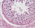
===Testis Histology=== | ===Spermatogonia=== | ||
* first cells of spermatogenesis. | |||
* originate in week 4 of fetal development in the endodermal walls of the yolk sac and migrate to the primordium of the testis, where they differentiate into spermatogonia. | |||
* Spermatogonia remain dormant until puberty. They are always in contact with the basal lamina of the tubule. | |||
* Two types of spermatogonia can be distinguished in the human seminiferous epithelium: | |||
** Type A spermatogonia have a rounded nucleus with very fine chromatin grains and one or two nucleoli. They are stem cells which divide to form new generations of both type A and type B spermatogonia. | |||
** Type B spermatogonia have rounded nuclei with chromatin granules of variable size, which often attach to the nuclear membrane, and one nucleolus. Although type B spermatogonia may divide repeatedly, they do not function as stem cells and their final mitosis always results in the formation of primary spermatocytes. | |||
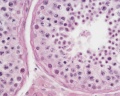
===Primary spermatocytes=== | |||
* lie in the cell layer luminal to the spermatogonia. | |||
* appear larger than spermatogonia. | |||
* immediately enter the prophase of the first meiotic division, which is prolonged (about 22 days). | |||
* large number of primary spermatocytes is always visible in cross-sections through seminiferous tubules. | |||
* cell divisions, from the formation of primary spermatocytes and onwards, to the production of the spermatocytes, are incomplete. | |||
* cells remain connected by bridges of cytoplasm. | |||
* completion of the first meiotic division results in the formation of secondary spermatocytes | |||
===Secondary spermatocytes=== | |||
* smaller than primary spermatocytes. | |||
* rapidly enter and complete the second meiotic division (seldom seen in histological preparations). | |||
* division results in the formation of spermatids. | |||
===Spermatids=== | |||
* lie in the luminal part of the seminiferous epithelium. | |||
* small cells (about 10 µm in diameter) with an initially very light (often eccentric) nucleus. | |||
** chromatin condenses during the maturation of the spermatids into spermatozoa, and the nucleus becomes smaller and stains darker. | |||
* terminal phase of spermatogenesis is called spermiogenesis | |||
** consists of the morphological differentiation of the newly formed spermatids into spermatozoa. | |||
| [[File:Testis histology 1.jpg]] | |||
|- | |||
|[[File:Seminiferous-tubule-HEx40.jpg]] | |||
| [[File:Testis_histology_2.jpg]] | |||
|- | |||
|} | |||
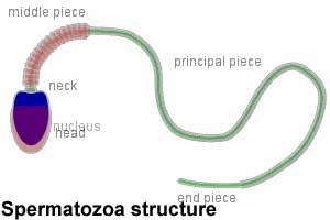
==Spermatozoa== | |||
{| | |||
| [[File:spermatozoa animation icon.jpg|100px]] This brief animation shows an overview of the structural components of the [[S#spermatozoa|spermatozoa]]. | |||
# Blue - Nucleus containing male haploid genome required to combine with oocyte haploid genome to form diploid zygote. | |||
# Red - Acrosome containing enzymes required to digest the zona pellucida. The acrosome develops as a highly modified golgi structure. | |||
# Green - Centriole and axonema required for spermatozoa movement. | |||
# Orange - Mitochondria and annulus required for energy for spermatozoa movement. | |||
# Grey - Cytoplasm and plasmolemma, cell membrane containing membrane proteins for chemotaxis and binding to the oocyte zone pellucida. | |||
* mature human spermatozoon is about 60 µm long and actively motile. | |||
* divided into head, neck and tail. | |||
** head - (flattened, about 5 µm long and 3 µm wide) chiefly consists of the nucleus (greatly condensed chromatin!). The anterior 2/3 of the nucleus is covered by the acrosome, which contains enzymes important in the process of fertilisation. The posterior parts of the nuclear membrane forms the so-called basal plate. | |||
** neck - short (about 1 µm) and attached to the basal plate. A transversely oriented centriole is located immediately behind the basal plate. The neck also contains nine segmented columns of fibrous material, which continue as the outer dense fibres into the tail.
| |||
** tail - further divided into a middle piece, a principal piece and an end piece. The '''axonema''' (arrangement of microtubules in all cilia) begins in the middle piece. It is surrounded by nine outer dense fibres, which are not found in other cilia. In the middle piece (about 5 µm long), the axonema and dense fibres are surrounded by a sheath of mitochondria. The middle piece is terminated by a dense ring, the annulus. The principal piece is about 45 µm long. It contains a fibrous sheath, which consists of dorsal and ventral longitudinal columns interconnected by regularly spaced circumferential hoops. The fibrous sheath and the dense fibres do not extend to the tip of the tail. Along the last part (5 µm) of the tail, called the end piece, the axonema is only surrounded by a small amount of cytoplasm and the plasma membrane. | |||
| <mediaplayer width='300' height='240' image="http://embryology.med.unsw.edu.au/embryology/images/0/02/Spermatozoa_animation_icon.jpg">File:Spermatozoa_animation.mp4</mediaplayer> | |||
|} | |||
<gallery> | |||
File:Spermatozoa_histology_001.jpg|x20 | |||
File:Spermatozoa_histology_002.jpg|x40 | |||
File:Spermatozoa_histology_003.jpg|x100 | |||
</gallery> | |||
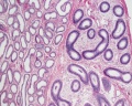
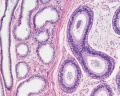
==Epididymis Histology== | |||
{| | |||
| Pseudostratified Epithelium | |||
* Nuclei of the epithelial cells are typically located in the widest part of the cell. | |||
** the nuclei of cells that do or do not reach the surface of the epithelium are often located at different heights within the epithelium, giving the epithelium a "stratified" appearance. | |||
* The epithelium though is not stratified, therefore named "pseudostratified". | |||
| [[File:Testis_histology_023.jpg]] | |||
|- | |||
| [[File:Epididymis histology 02.jpg]] | |||
| [[File:Epididymis histology 03.jpg]] | |||
|} | |||
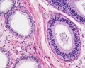
==Human Testis (adult)== | |||
<gallery> | |||
File:Testis_histology_002.jpg|overview x2 | |||
File:Testis_histology_010.jpg|convoluted seminiferous tubules x10 | |||
File:Testis_histology_009.jpg|convoluted seminiferous tubules x20 | |||
File:Testis_histology_011.jpg|convoluted seminiferous tubules x40 | |||
File:Testis_histology_012.jpg|convoluted seminiferous tubules x40 | |||
File:Testis_histology_008.jpg|epididymis overview x4 | |||
File:Testis_histology_013.jpg|epididymis x10 | |||
File:Testis_histology_014.jpg|epididymis x20 | |||
File:Testis_histology_015.jpg|epididymis x40 | |||
File:Testis_histology_003.jpg|ductus deferens overview x2 | |||
File:Testis_histology_016.jpg|ductus deferens x10 | |||
File:Testis_histology_017.jpg|ductus deferens x40 | |||
</gallery> | |||
==Human Testis (young)== | |||
<gallery> | |||
File:Testis_histology_001.jpg|overview Loupe | |||
File:Testis_histology_004.jpg|convoluted seminiferous tubules x10 | |||
File:Testis_histology_006.jpg|convoluted seminiferous tubules x40 | |||
File:Testis_histology_007.jpg|convoluted seminiferous tubules x40 | |||
File:Testis_histology_005.jpg|tunica albuginea x20 | |||
</gallery> | |||
{{Testis Histology}} | {{Testis Histology}} | ||
==Other Species== | |||
===Rabbit=== | |||
<gallery> | |||
File:Testis_histology_018.jpg|convoluted seminiferous tubules x20 | |||
File:Testis_histology_019.jpg|convoluted seminiferous tubules x100 | |||
</gallery> | |||
===Mouse=== | |||
<gallery> | |||
File:Mouse epididymis development 01.jpg|postnatal epididymis | |||
File:Mouse epididymis development 02.jpg|14 days postnatal | |||
File:Mouse epididymis development 03.jpg|33 days postnatal | |||
File:Mouse epididymis development 04.jpg|45 days postnatal | |||
File:Mouse epididymis development 05.jpg|2 months postnatal | |||
</gallery> | |||
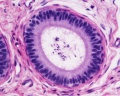
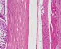
==Ductus Deferens Histology== | |||
[[File:Ductus_deferens_01.jpg]][[File:Ductus_deferens_02.jpg]] | |||
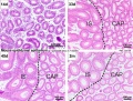
==Prostate Histology== | |||
{| | |||
| [[File:Prostate_histology_01.jpg|300px]] | |||
| [[File:Prostate_histology_02.jpg|300px]] | |||
| [[File:Prostate_histology_03.jpg|300px]] | |||
|- | |||
| '''Human prostate histology''' | |||
| '''Corpora Amylacea''' | |||
| '''Submucosal gland''' | |||
|- | |||
| (adult, low power overview) | |||
| (adult, detail) | |||
| (adult, high power detail) | |||
|} | |||
==Penis Histology== | |||
<gallery> | |||
File:Male_histology_001.jpg| | |||
File:Male_histology_002.jpg| | |||
File:Male_histology_003.jpg| | |||
File:Male_histology_004.jpg| | |||
</gallery> | |||
==Terms== | |||
* '''cortex''' - (Latin = rind, or bark) outer layer of an organ. | |||
* '''hilum''' - or hilus (Latin,= a trifle; depression in a seed) a depression at vascular entrance/exit of a gland or organ. | |||
* '''medulla''' - (Latin, ''medulla'' = pith, marrow) the inner portion of an organ, in contrast to cortex. | |||
* '''mucosa''' - (Latin, = mucous membrane) thin layer which lines body cavities and passages formed by epithelium and lamina propria. | |||
* '''parenchyma''' - (Greek," + enkeim = to pour in) the essential functional cells of an organ as opposed to its stroma. | |||
* '''serosa''' - (Latin, ''serum'' = whey; a pale fluid) a serous membrane lining body cavities. | |||
* '''stroma''' - (Greek, = a cover, table-cloth, bedding) term for the internal supporting frame-work of a tissue, or organ, as opposed to its parenchyma. | |||
* '''tunica albuginea''' - a dense, white, fibrous sheath enclosing a part or organ. | |||
---- | |||
:'''Links:''' [[Genital_-_Female_Development|Female]] | [[Ovary Development|Ovary]] | [[Oocyte Development|Oocyte]] | [[Uterus Development|Uterus]] | [[Vagina Development|Vagina]] | |||
{{Histology Glossary}} | |||
Revision as of 13:57, 1 June 2013

| ANAT2241 This practical support page content is not part of the virtual science practical class and provides additional information for student self-directed learning purposes. All practical class pages are located on Moodle - ANAT2241 |
General Objective
To know the histological and cytological structures of the major components of the male reproductive system.
Specific Objectives
- To describe the microanatomy of the testis and epididymis.
- To identify cells of the germinal epithelium of the seminiferous tubule: Sertoli cells, spermatogonia, spermatocytes, spermatids and spermatozoa.
- To know the main events occurring in spermiogenesis.
- To know the structure of the ductus deferens, seminal vesicle, prostate gland and penis.
Learning Activities
Examine the following virtual slides, and in course manual identify, draw and label the structures and note their function.
Virtual Slides: ANAT2241 Male Reproductive
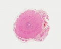
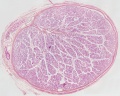
Testis Histology
Convoluted Seminiferous Tubules
Spermatogonia
** Type A spermatogonia have a rounded nucleus with very fine chromatin grains and one or two nucleoli. They are stem cells which divide to form new generations of both type A and type B spermatogonia. ** Type B spermatogonia have rounded nuclei with chromatin granules of variable size, which often attach to the nuclear membrane, and one nucleolus. Although type B spermatogonia may divide repeatedly, they do not function as stem cells and their final mitosis always results in the formation of primary spermatocytes. Primary spermatocytes
Secondary spermatocytes
Spermatids
|

|

|

|
Spermatozoa
|
<mediaplayer width='300' height='240' image="http://embryology.med.unsw.edu.au/embryology/images/0/02/Spermatozoa_animation_icon.jpg">File:Spermatozoa_animation.mp4</mediaplayer> |
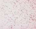
x20
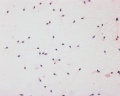
x40
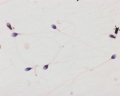
x100


Epididymis Histology
Pseudostratified Epithelium
|

|

|

|
Human Testis (adult)
overview x2
convoluted seminiferous tubules x10
convoluted seminiferous tubules x20
convoluted seminiferous tubules x40
convoluted seminiferous tubules x40
epididymis overview x4
epididymis x10
epididymis x20
epididymis x40
ductus deferens overview x2

ductus deferens x10
ductus deferens x40


Human Testis (young)
overview Loupe
convoluted seminiferous tubules x10

convoluted seminiferous tubules x40
convoluted seminiferous tubules x40
tunica albuginea x20


Testis Histology Links: Testis Development | Spermatozoa Development | Histology
- Human (young): overview labeled | overview unlabeled | convoluted seminiferous tubules x10 | x40 | x40 | tunica albuginea x20
- Human (adult): overview x2 | convoluted seminiferous tubules labeled | x10 | x20 | x40 | x40 | epididymis ductulus efferens | ductus epididymidis | epithelium | overview x4 | x10 | x20 | x40 | ductus deferens labeled overview | epithelium | overview x2 | x10 | x40
- Human Stage 22: Testis - labeled overview | Testis - unlabeled overview | Testis - unlabeled detail | Testis - labeled detail | testis | Carnegie stage 22 | Movie - Urogenital stage 22
- Mouse: postnatal epididymis | 14 days postnatal | 33 days postnatal | 45 days postnatal | 2 months postnatal
| Spermatozoa Development (expand to see terms) | ||
|---|---|---|
|
Note there are additional glossaries associated with genital, spermatozoa, oocyte and renal.
See also: Spermatozoa Terms collapse table
|

Other Species
Rabbit

convoluted seminiferous tubules x20

convoluted seminiferous tubules x100


Mouse
postnatal epididymis
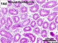
14 days postnatal
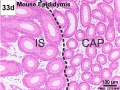
33 days postnatal
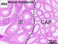
45 days postnatal
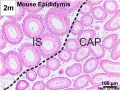
2 months postnatal


Ductus Deferens Histology


Prostate Histology

|

|

|
| Human prostate histology | Corpora Amylacea | Submucosal gland |
| (adult, low power overview) | (adult, detail) | (adult, high power detail) |
Penis Histology


Terms
- cortex - (Latin = rind, or bark) outer layer of an organ.
- hilum - or hilus (Latin,= a trifle; depression in a seed) a depression at vascular entrance/exit of a gland or organ.
- medulla - (Latin, medulla = pith, marrow) the inner portion of an organ, in contrast to cortex.
- mucosa - (Latin, = mucous membrane) thin layer which lines body cavities and passages formed by epithelium and lamina propria.
- parenchyma - (Greek," + enkeim = to pour in) the essential functional cells of an organ as opposed to its stroma.
- serosa - (Latin, serum = whey; a pale fluid) a serous membrane lining body cavities.
- stroma - (Greek, = a cover, table-cloth, bedding) term for the internal supporting frame-work of a tissue, or organ, as opposed to its parenchyma.
- tunica albuginea - a dense, white, fibrous sheath enclosing a part or organ.
- Histology Glossary: A | B | C | D | E | F | G | H | I | J | K | L | M | N | O | P | Q | R | S | T | U | V | W | X | Y | Z | ANAT2241 Support | Histology | Histology Stains | Embryology Glossary
Course Links
- Histology Glossary: A | B | C | D | E | F | G | H | I | J | K | L | M | N | O | P | Q | R | S | T | U | V | W | X | Y | Z | ANAT2241 Support | Histology | Histology Stains | Embryology Glossary
| Common Histology Stains | ||||||||||||||||||||||||||||||||||||||||||||||||||||||||||||||||||||||||||||||||||||||||||||||||||||||||||||||||||||||||||||||||||||||||||||||||
|---|---|---|---|---|---|---|---|---|---|---|---|---|---|---|---|---|---|---|---|---|---|---|---|---|---|---|---|---|---|---|---|---|---|---|---|---|---|---|---|---|---|---|---|---|---|---|---|---|---|---|---|---|---|---|---|---|---|---|---|---|---|---|---|---|---|---|---|---|---|---|---|---|---|---|---|---|---|---|---|---|---|---|---|---|---|---|---|---|---|---|---|---|---|---|---|---|---|---|---|---|---|---|---|---|---|---|---|---|---|---|---|---|---|---|---|---|---|---|---|---|---|---|---|---|---|---|---|---|---|---|---|---|---|---|---|---|---|---|---|---|---|---|---|---|
| ||||||||||||||||||||||||||||||||||||||||||||||||||||||||||||||||||||||||||||||||||||||||||||||||||||||||||||||||||||||||||||||||||||||||||||||||
| ||||||||||||||||||||||||||||||||||||||||||||||||||||||||||||||||||||||||||||||||||||||||||||||||||||||||||||||||||||||||||||||||||||||||||||||||
Practical Support
- Pages can be accessed from any internet connected computer.
ANAT2241 Support Links: The Virtual Microscope | Covering and Lining Epithelia | Glandular Epithelia | CT Components | CT Types | Bone, Bone Formation and Joints | Muscle | Nervous | Blood | Eye | Cardiovascular | Respiratory | Integumentary | Gastrointestinal | Gastrointestinal Organs | Lymphatic and Immune | Endocrine | Urinary | Female Reproductive | Male Reproductive | Histology Stains | Histology Drawings | Practicals Health and Safety 2013 | Moodle - 2019
ANAT2241 This practical support page content is not part of the science practical class and provides only background information for student self-directed learning purposes.
Cite this page: Hill, M.A. (2024, June 10) Embryology ANAT2241 Male Reproductive System. Retrieved from https://embryology.med.unsw.edu.au/embryology/index.php/ANAT2241_Male_Reproductive_System
- © Dr Mark Hill 2024, UNSW Embryology ISBN: 978 0 7334 2609 4 - UNSW CRICOS Provider Code No. 00098G