File:Spleen histology 01.jpg: Difference between revisions
From Embryology
No edit summary |
No edit summary |
||
| Line 2: | Line 2: | ||
'''White Pulp''' | '''White Pulp''' | ||
* lymphocytes surround central | * central arteriole | ||
* lymphocytes surround central arteriole as periarterial lymphoid sheath (PALS) | |||
* B and T cells | |||
'''Red Pulp''' | '''Red Pulp''' | ||
| Line 9: | Line 11: | ||
* Splenic cords | * Splenic cords | ||
'''Marginal Zone''' | |||
* white and red pulp interact | |||
No real cortex and medulla. | No real cortex and medulla. | ||
Revision as of 08:53, 27 February 2012
Spleen Histology - Overview Red and White Pulp
White Pulp
- central arteriole
- lymphocytes surround central arteriole as periarterial lymphoid sheath (PALS)
- B and T cells
Red Pulp
- Red blood cells
- Splenic sinuses
- Splenic cords
Marginal Zone
- white and red pulp interact
No real cortex and medulla.
Links: Histology | Histology Stains | Blue Histology images copyright Lutz Slomianka 1998-2009. The literary and artistic works on the original Blue Histology website may be reproduced, adapted, published and distributed for non-commercial purposes. See also the page Histology Stains.
Cite this page: Hill, M.A. (2024, May 23) Embryology Spleen histology 01.jpg. Retrieved from https://embryology.med.unsw.edu.au/embryology/index.php/File:Spleen_histology_01.jpg
- © Dr Mark Hill 2024, UNSW Embryology ISBN: 978 0 7334 2609 4 - UNSW CRICOS Provider Code No. 00098G
Original File name: Spl041he.jpg
File history
Click on a date/time to view the file as it appeared at that time.
| Date/Time | Thumbnail | Dimensions | User | Comment | |
|---|---|---|---|---|---|
| current | 19:26, 22 February 2012 | 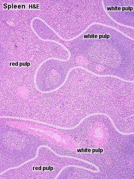 | 450 × 600 (133 KB) | Z8600021 (talk | contribs) | |
| 14:53, 21 February 2011 | 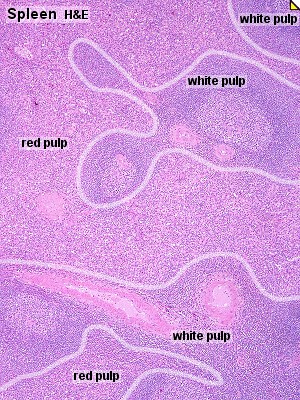 | 300 × 400 (73 KB) | S8600021 (talk | contribs) | ==Spleen Histology== Original File name: Spl041he.jpg {{Blue Histology}} Category:Spleen Category:Endocrine Category:Histology Category:Immune |
You cannot overwrite this file.