File:Human embryo tomography Carnegie stage 17.jpg: Difference between revisions
From Embryology
(==Human Embryo Carnegie Stage 17 Optical Projection Tomography (OPT) Model== * The embryo was staged as CS17, which is approximately 41 days of development. * The developing central nervous system (CNS) is clearly visible even in the external view of th) |
|||
| Line 6: | Line 6: | ||
* There is very little detail in the developing liver compared to the CNS. | * There is very little detail in the developing liver compared to the CNS. | ||
* Blood vessels, dorsal root ganglia and developing vertebrae are clearly visible. | * Blood vessels, dorsal root ganglia and developing vertebrae are clearly visible. | ||
:'''Links:''' [[Carnegie stage 17]] | [[Quicktime Movie Carnegie stage 17|Quicktime]] | [[Movie Carnegie stage 17|Flash]] | |||
===Reference=== | ===Reference=== | ||
<pubmed>15298700</pubmed>| [http://www.ncbi.nlm.nih.gov/pmc/articles/PMC514604 PMC514604] | [http://www.biomedcentral.com/1471-2202/5/27 BMC Neurosci.] | <pubmed>15298700</pubmed>| [http://www.ncbi.nlm.nih.gov/pmc/articles/PMC514604 PMC514604] | [http://www.biomedcentral.com/1471-2202/5/27 BMC Neurosci.] | ||
[http://www.ncl.ac.uk/ihg/EADHB Electronic Atlas of the Developing Human Brain] | |||
Revision as of 01:17, 21 February 2012
Human Embryo Carnegie Stage 17 Optical Projection Tomography (OPT) Model
- The embryo was staged as CS17, which is approximately 41 days of development.
- The developing central nervous system (CNS) is clearly visible even in the external view of the CS17 model.
- Differences in autofluorescence within the CNS and among different organs are also apparent.
- There is very little detail in the developing liver compared to the CNS.
- Blood vessels, dorsal root ganglia and developing vertebrae are clearly visible.
- Links: Carnegie stage 17 | Quicktime | Flash
Reference
<pubmed>15298700</pubmed>| PMC514604 | BMC Neurosci.
Electronic Atlas of the Developing Human Brain
© 2004 Kerwin et al; licensee BioMed Central Ltd.
This is an open-access article distributed under the terms of the Creative Commons Attribution License (http://creativecommons.org/licenses/by/2.0), which permits unrestricted use, distribution, and reproduction in any medium, provided the original work is properly cited.
CS17 OPT model.mpg. Mpeg movie of 3D CS17 OPT model. Human embryo tomography Carnegie stage 17.mov
Human embryo tomography Carnegie stage 17.flv
Original file name 1471-2202-5-27-s1.mpg
File history
Click on a date/time to view the file as it appeared at that time.
| Date/Time | Thumbnail | Dimensions | User | Comment | |
|---|---|---|---|---|---|
| current | 01:06, 10 March 2012 |  | 516 × 516 (35 KB) | Z8600021 (talk | contribs) | added week 6 label |
| 01:05, 10 March 2012 | 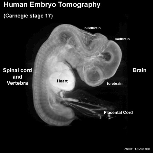 | 516 × 516 (34 KB) | Z8600021 (talk | contribs) | added additional labels | |
| 01:03, 21 February 2012 |  | 516 × 516 (28 KB) | S8600021 (talk | contribs) | ==Human Embryo Carnegie Stage 17 Optical Projection Tomography (OPT) Model== * The embryo was staged as CS17, which is approximately 41 days of development. * The developing central nervous system (CNS) is clearly visible even in the external view of th |
You cannot overwrite this file.
File usage
The following 19 pages use this file:
- 2017BGDLecture-Neural-Movie
- BGDA Lecture - Development of the Nervous System
- Brain Awareness Week 2012
- Carnegie Stage 17 Neural Movie
- Carnegie stage 17
- Computed Tomography
- Ectoderm
- HDBR and HuDSeN Collection
- Human Embryo Collections
- K12 Brain Awareness Week
- Movie - Carnegie Stage 17 Neural
- Movies
- Neural System Development
- Quicktime Movie - Carnegie Stage 17 Neural
- Talk:Carnegie Stage 17 Neural Movie
- Talk:Flash Movies
- Talk:K12 Brain Awareness Week
- Template:Neural cartoons
- Template:Stage 17 Embryo movie