File:Boyd1950 fig01.jpg: Difference between revisions
From Embryology
(===Reference=== {{Ref-Boyd1950}}) |
mNo edit summary |
||
| Line 1: | Line 1: | ||
==Fig. 1. Transverse section through right third pouch of a 10 mm human embryo== | |||
(X 400). Parathyroid tissue already shows clear signs of differentiation. The cervical vesicle IV has separated from the surface endoderm of the region of the cervical sinus. | |||
===Reference=== | ===Reference=== | ||
{{Ref-Boyd1950}} | {{Ref-Boyd1950}} | ||
{{Footer}} | |||
Revision as of 21:20, 6 March 2017
Fig. 1. Transverse section through right third pouch of a 10 mm human embryo
(X 400). Parathyroid tissue already shows clear signs of differentiation. The cervical vesicle IV has separated from the surface endoderm of the region of the cervical sinus.
Reference
Boyd JD. Development of the thyroid and parathyroid glands and the thymus. (1950) Ann R Coll Surg Engl. 7(6): 455-71. PMID 14790564
Cite this page: Hill, M.A. (2024, June 3) Embryology Boyd1950 fig01.jpg. Retrieved from https://embryology.med.unsw.edu.au/embryology/index.php/File:Boyd1950_fig01.jpg
- © Dr Mark Hill 2024, UNSW Embryology ISBN: 978 0 7334 2609 4 - UNSW CRICOS Provider Code No. 00098G
File history
Click on a date/time to view the file as it appeared at that time.
| Date/Time | Thumbnail | Dimensions | User | Comment | |
|---|---|---|---|---|---|
| current | 21:21, 6 March 2017 | 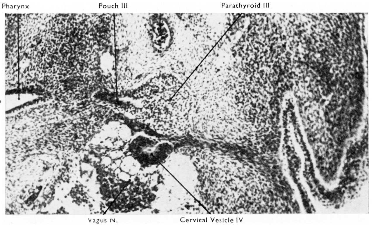 | 1,280 × 769 (247 KB) | Z8600021 (talk | contribs) | |
| 21:20, 6 March 2017 |  | 1,320 × 919 (380 KB) | Z8600021 (talk | contribs) | ===Reference=== {{Ref-Boyd1950}} |
You cannot overwrite this file.
File usage
The following page uses this file: