Category:2011 Student Image: Difference between revisions
From Embryology
m (Protected "Category:2011 Student Image": Protect from student change ([edit=sysop] (indefinite) [move=sysop] (indefinite))) |
No edit summary |
||
| Line 4: | Line 4: | ||
If image reuse on this non-commercial educational site infringes your existing copyright, please [mailto:m.hill@unsw.edu.au contact the site editor] for immediate removal. | If image reuse on this non-commercial educational site infringes your existing copyright, please [mailto:m.hill@unsw.edu.au contact the site editor] for immediate removal. | ||
'''Links:''' [[Science]] | |||
[[Category:Science-Undergraduate]] [[Category:2011] | |||
Latest revision as of 11:55, 14 July 2012
The images shown below were originally uploaded as part of a 2011 student project and may contain inaccuracies in either description or acknowledgements.
Students have been advised in writing concerning the reuse of content and may accidentally have misunderstood the original terms of use.
If image reuse on this non-commercial educational site infringes your existing copyright, please contact the site editor for immediate removal.
Links: Science [[Category:2011]
Pages in category '2011 Student Image'
The following 5 pages are in this category, out of 5 total.
Media in category '2011 Student Image'
The following 193 files are in this category, out of 193 total.
 22+23=45.jpg 594 × 158; 11 KB
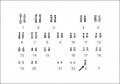
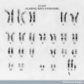 47,XXY Klinefelter's Syndrome.jpg 576 × 576; 58 KB
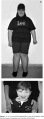 A 12 year old PWS patient and a 4 year old AS patient.jpg 358 × 966; 141 KB
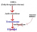 Action of Amoratase Inhibitors on Production of Estradiol.JPG 316 × 276; 12 KB
 Angelman Syndrome patient.png 343 × 271; 88 KB
 Angelo DiGeorge.png 331 × 480; 170 KB
 Angiography image indicating Supravalvular aortic stenosis.jpg 788 × 577; 133 KB
 Bilateral Cleft Lip Variations.jpg 619 × 714; 87 KB
 Bilateral cleft lip with cleft hard and soft palate.jpg 168 × 137; 5 KB
 Bilateral cleft lip with cleft hard palate.jpg 167 × 141; 5 KB
 Bilateral Cleft Lip With Nasal Deformity.jpg 685 × 539; 148 KB
 Bilateral cleft lip.jpg 169 × 137; 5 KB
 Blood Loss During Lip and Palate Repair.jpg 600 × 606; 21 KB
 Blood test result for glucose and iron.jpg 879 × 345; 54 KB
 Blood test results.jpg 879 × 345; 54 KB
 Chromosome 20 - JAG1 gene.jpg 976 × 552; 55 KB
 Chromosome 22 - TBX1 Gene.jpg 960 × 540; 71 KB
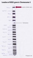
 Chromosome 5 - NKX2-5 Gene.jpg 960 × 540; 70 KB
 Chromosome 7, indicating 7q11.23 region of Williams Syndrome.gif 325 × 270; 11 KB
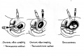
 Cleft Palate Maxillary and Mandibular View.jpg 547 × 658; 141 KB
 Cognitive performance in WS subjects (n = 67) versus normal controls.png 600 × 292; 139 KB
 Comparison of morphogenesis of the upper lip with the palate.jpg 774 × 371; 188 KB
 Control group response to startle.jpg 661 × 492; 66 KB
 Critical region of Angelman Syndrome on chromosome 15.png 591 × 576; 356 KB
 Cross Section of the Spinal Cord.jpg 1,280 × 640; 74 KB
 CT lungcancer.JPG 422 × 348; 18 KB
 Cyanotic baby.jpg 469 × 600; 43 KB
 Development 2.jpg 588 × 637; 22 KB
 Development 3.jpg 588 × 637; 21 KB
 Development1.jpg 588 × 637; 22 KB
 DiGeorge Baby.jpg 691 × 800; 64 KB
 Dr Charles Williams.jpg 430 × 500; 101 KB
 Dr Etienne Louis Arthur Fallot.jpg 434 × 576; 53 KB
 Dr Harry Angelman.jpg 481 × 600; 49 KB
 Dr Helen Brooke Taussig.jpg 307 × 418; 29 KB
 Duchenne.JPG 324 × 457; 22 KB
 Dystrophin in the muscle fibre membrane.jpg 1,840 × 1,355; 415 KB
 Dystrophin within the plasma membrane of muscle fibres.jpg 800 × 640; 112 KB
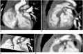
 Echocardiogram concentric left ventricular hypertrophy.jpg 800 × 460; 55 KB
 Effect of Frataxin Levels.jpg 1,360 × 624; 84 KB
 Electrocardiograph findings in dogs affected with DMD.JPG 609 × 538; 64 KB
 Electroencephalography of Angelman Syndrome.jpg 440 × 207; 33 KB
 Emotional Response to 'cartoon' test Klinefelter Syndrome.png 1,008 × 630; 174 KB
 Extent of microcephaly in Angelman Syndrome patients.png 1,000 × 814; 43 KB
 Facial features of four individuals with Willams Syndrome.gif 429 × 182; 45 KB
 Finger-Clubbing.jpg 600 × 449; 58 KB
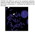 FISH test used to confirm the deletion of the ELN gene.jpg 337 × 312; 15 KB
 Fluorescence In situ Hybridization (FISH) assay.JPG 696 × 222; 11 KB
 FMR1 is silenced in FXS.jpg 451 × 497; 21 KB
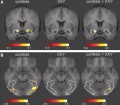 FMRI Images of Brain Activation in XXY Patients.JPG 433 × 378; 23 KB
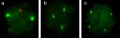
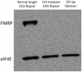 FMRP expression in control and fragile X tissues.png 600 × 527; 1.29 MB
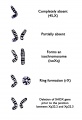
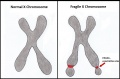 Fragile X Chromosome..jpg 997 × 656; 69 KB
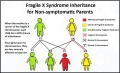
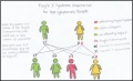 Fragile x inheritance..jpg 1,074 × 654; 66 KB
 Fragile X Inheritance..jpg 1,074 × 654; 105 KB
 Fragile x inheritance.jpg 1,074 × 654; 102 KB
 Fragile X Phenotype.jpg 225 × 112; 23 KB
 Friedreich's Ataxia Pedigree.jpg 1,424 × 588; 80 KB
 Friedreich's Ataxia Pedigree.png 618 × 384; 58 KB
 From infancy until completion of treatment.jpg 702 × 254; 103 KB
 Furlow Z-plasty technique.jpg 670 × 771; 147 KB
 GAA Frequency in FRDA.jpg 622 × 345; 37 KB
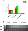
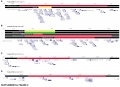
 Gene expression responses of Friedreich's ataxia.jpg 551 × 749; 342 KB
 Genotyping of the five microsatellites markers in WBS families.jpg 465 × 213; 71 KB
 George Huntington.jpg 285 × 358; 44 KB
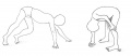 Gower's sign - a symptom of DMD.JPG 1,144 × 491; 36 KB
 HD future research.jpg 687 × 869; 78 KB
 HD Interview questions.png 204 × 135; 12 KB
 HD patient with no treatment.mov ; 1.17 MB
 HD patients.jpg 492 × 442; 52 KB
 Heart disection.jpg 963 × 731; 209 KB
 Heart Hypertrophy gross.jpg 639 × 800; 59 KB
 House drawings Williams.jpg 2,328 × 1,464; 112 KB
 Huntingtin gene.jpeg 492 × 181; 15 KB
 Huntington disease atrophy 1.jpg 630 × 630; 148 KB
 Huntington disease atrophy 2.jpg 630 × 630; 146 KB
 Huntington disease atrophy 3.jpg 630 × 630; 144 KB
 Huntington Disease patient and control MRI.gif 530 × 352; 139 KB
 Huntington's disease MRI.jpg 504 × 630; 163 KB
 Imprint defect inheritance in Angelman Syndrome.png 688 × 323; 21 KB
 Inheritance pattern in Huntington's Disease.jpeg 531 × 284; 15 KB
 Introduction 3.jpg 921 × 637; 40 KB
 Introduction 4.jpg 907 × 637; 31 KB
 JAG1.jpeg 196 × 139; 6 KB
 Karyotype of a Klinefelter's syndrome patient.jpg 779 × 545; 85 KB
 Karyotype of Klinefelter's Syndrome.png 768 × 600; 222 KB
 Karyotype.jpg 648 × 454; 49 KB
 Key cellular pathogenic mechanisms in HD.jpg 663 × 517; 47 KB
 Key cellular pathogenic mechanisms in Huntington's disease.jpg 663 × 517; 47 KB
 Klinefelter syndrome group response to startle.jpg 661 × 492; 73 KB
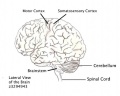 Lateral View of the Brain.jpeg 782 × 627; 102 KB
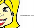 Lowered Ear Lobes.jpg 1,078 × 828; 72 KB
 Major causes of death in surgically untreated TOF patients.png 1,009 × 570; 127 KB
 Maternal Non-Disjunction.PNG 952 × 525; 52 KB
 Mechanism of tetrabenazine.jpg 600 × 459; 33 KB
 Median facial dysplasia.jpg 597 × 448; 179 KB
 Meiotic non-disjunction.jpg 989 × 299; 35 KB
 Mice mutants exhibit cleft palate and umbilical hernia.jpg 527 × 332; 126 KB
 Migration.jpg 771 × 333; 118 KB
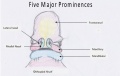 Modified prominences final.jpg 667 × 423; 23 KB
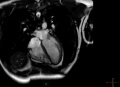 MRI heart.JPG 491 × 354; 14 KB
 MSX 1 Gene.JPG 385 × 677; 22 KB
 Neuroacanthocytosis.jpg 487 × 584; 91 KB
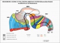 NeuromericOrganization.jpg 620 × 449; 123 KB
 Nikolaus Friedreich Portrait.jpg 320 × 576; 54 KB
 NKX2-5.jpeg 469 × 173; 14 KB
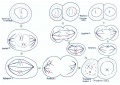 Nondisjunction of Homologous Chromosomes in Meiosis1.jpg 900 × 636; 113 KB
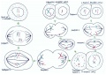 Nondisjunction of Sister Chromatids in Meiosis 2.jpg 900 × 636; 121 KB
 Nondisjunction.jpg 638 × 135; 23 KB
 Normal and Angelman Syndrome mice models.jpg 700 × 505; 135 KB
 Normal fetal blood flow and Tetralogy of Fallot.jpg 628 × 543; 200 KB
 Normal palate shelf and key stages of mouse palatal development.jpg 771 × 153; 80 KB
 Oral Clefting.JPG 662 × 464; 31 KB
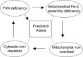
 Pathogenesis of Friedreich Ataxia.jpg 520 × 380; 30 KB
 Pes Cavus Deformity.jpg 649 × 247; 79 KB
 Phenotypes of FXS overlap with those of Autism.jpg 600 × 441; 51 KB
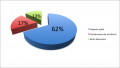
 Pie Chart.JPG 551 × 572; 50 KB
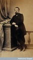
 Pierre Joseph Desault.png 1,431 × 2,100; 2.34 MB
 Point mutations resulting in DMD.jpg 2,051 × 1,131; 412 KB
 Point vs frameshift mutation of DMD gene.png 1,689 × 1,000; 874 KB
 Potts Shunt.jpg 311 × 456; 117 KB
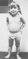
 Prader-Willi Syndrome patient.png 343 × 650; 149 KB
 Psoriasis.jpg 275 × 181; 9 KB
 Pubertal gynecomastia 1.jpg 550 × 364; 92 KB
 Regions of the brain significant in Huntington's disease.jpg 1,280 × 1,239; 348 KB
 Regions of the brain.jpg 1,280 × 1,239; 348 KB
 Repair of Bilateral Cleft Lip.jpg 618 × 459; 179 KB
 Role of FXN Gene.jpg 514 × 356; 51 KB
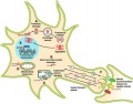
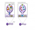 Role of UBE3A in dendritic spine neuronal synapses.png 725 × 621; 94 KB
 Scans of Supravalvular Aortic Stenosis and Pulmonary Stenosis.jpg 665 × 436; 114 KB
 Schematic diagram detailing genes deleted in probands.jpeg 2,850 × 2,075; 480 KB
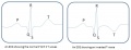 Schematic ECG normal and inverted T-wave.jpg 1,001 × 384; 32 KB
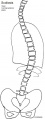 Scoliosis drawn.jpg 404 × 1,070; 43 KB
 Speckle Tracking Echocardiograph of a dog affected with DMD.JPG 651 × 441; 67 KB
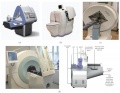 SPECT scanner.jpg 594 × 462; 161 KB
 Spinal problems DMD.jpg 568 × 376; 127 KB
 Stats abnormal.jpg 392 × 263; 32 KB
 Subtle Finger tremors-Angelman Syndrome.jpg 600 × 454; 82 KB
 Symptoms and signs in FRDA patients.jpg 700 × 412; 52 KB
 Symptoms of DMD.JPG 626 × 676; 39 KB
 Symptoms of Fragile X Syndrome.JPG 576 × 470; 25 KB
 TBX1 gene.jpg 960 × 540; 50 KB
 TBX1.jpeg 181 × 185; 6 KB
 Tetrabenazine structure.JPG 600 × 450; 22 KB
 Tetralogy of fallot-the 4 defects.jpg 311 × 456; 42 KB
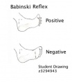 The Babinski Reflex.jpg 386 × 443; 25 KB
 The Blalock Taussig Shunt.jpg 466 × 684; 60 KB
 The frataxin gene on chromosome 9.jpg 689 × 909; 40 KB
 The Glenn Shunt.jpg 311 × 456; 125 KB
 Timeline of Cleft Lip & Palate development.png 1,374 × 181; 30 KB
 Tinsley et al. utrophin results.jpg 452 × 449; 114 KB
 Transverse section of the Cerebellum.jpg 1,016 × 629; 83 KB
 Turner Syndrome Test Showing Cystic Hygroma.gif 499 × 169; 70 KB
 Turner Syndrome Test Showing Nuchal Translucency.gif 453 × 254; 33 KB
 Turner Syndrome X Chromosome Variations.jpg 977 × 1,434; 130 KB
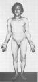 Turners syndrome 35 year old woman.jpg 444 × 943; 40 KB
 Turners syndrome 4 year old child.jpg 444 × 939; 40 KB
 TurnerSyndromeMaternalSerumSampling.jpg 1,589 × 1,059; 297 KB
 Type Bilateral cleft palate.jpg 166 × 144; 5 KB
 Type Unilateral Cleft Palate.jpg 164 × 143; 5 KB
 UBE3A Ubiquitylation Pathway.png 936 × 475; 168 KB
 Unilateral cleft lip with a cleft hard palate.jpg 166 × 137; 5 KB
 Unilateral cleft lip with cleft hard and soft palate.jpg 166 × 141; 5 KB
 Unilateral cleft lip.jpg 163 × 134; 5 KB
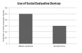 Use of social evaluation cues for WS and non WS individuals.jpg 2,396 × 1,552; 145 KB
 Van der Woude syndrome with lower lip pits.jpg 594 × 448; 170 KB
 Variations of Cleft Lip or Palate.jpg 951 × 1,050; 445 KB
 Velocardiofacial syndrome with typical facies.jpg 594 × 449; 171 KB
 Waterston Shunt.jpg 311 × 456; 119 KB
 Webbed Neck.jpg 299 × 234; 6 KB
 Williams Syndrome eye.jpg 1,198 × 766; 75 KB
 WS girl labelled.jpg 2,504 × 1,544; 246 KB
 X chromosome location of the dystrophin gene.jpg 876 × 1,521; 188 KB
 Z diagram Williams.jpg 2,328 × 1,464; 126 KB


_versus_normal_controls.png)


_vs._Duchennes_muscular_dystrophy_muscle_(b).jpg)


_assay.JPG)