File:Gall bladder histology 001.jpg: Difference between revisions
From Embryology
No edit summary |
No edit summary |
||
| Line 5: | Line 5: | ||
{{Gall Bladder Histology Images}} | {{Gall Bladder Histology Images}} | ||
{{Blue Histology}} | {{Blue Histology}} | ||
Original File name: Gall04he.jpg | |||
[[Category:Human]] [[Category:Gastrointestinal Tract]] [[Category:Histology]] | [[Category:Human]] [[Category:Gastrointestinal Tract]] [[Category:Histology]] | ||
Revision as of 10:17, 2 July 2012
Human Gall Bladder Histology
Gall bladder epithelium with underlying dense irregular connective tissue and then an irregular appearing muscular (smooth) layer. H&E
- Gallbladder Histology: overview (label) | overview (unlabel) | epithelium (label) | epithelium (unlabel) | GIT Histology
Links: Histology | Histology Stains | Blue Histology images copyright Lutz Slomianka 1998-2009. The literary and artistic works on the original Blue Histology website may be reproduced, adapted, published and distributed for non-commercial purposes. See also the page Histology Stains.
Cite this page: Hill, M.A. (2024, May 23) Embryology Gall bladder histology 001.jpg. Retrieved from https://embryology.med.unsw.edu.au/embryology/index.php/File:Gall_bladder_histology_001.jpg
- © Dr Mark Hill 2024, UNSW Embryology ISBN: 978 0 7334 2609 4 - UNSW CRICOS Provider Code No. 00098G
Original File name: Gall04he.jpg
File history
Click on a date/time to view the file as it appeared at that time.
| Date/Time | Thumbnail | Dimensions | User | Comment | |
|---|---|---|---|---|---|
| current | 01:40, 8 July 2011 | 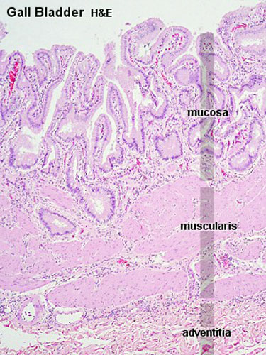 | 375 × 500 (78 KB) | S8600021 (talk | contribs) | |
| 01:40, 8 July 2011 | 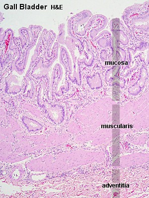 | 300 × 400 (65 KB) | S8600021 (talk | contribs) | ==Human Gall Bladder Histology== Showing central vein. H&E {{Liver Histology Images}} Original File name: Gall04he.jpg {{Blue Histology}} Category:Human Category:Gastrointestinal Tract Category:Histology |
You cannot overwrite this file.