File:Infant lymphocytic choriomeningitis virus CT.jpg: Difference between revisions
No edit summary |
|||
| Line 16: | Line 16: | ||
Journal.ppat.0030149.g001.jpg | Journal.ppat.0030149.g001.jpg | ||
[[Category:Human]] [[Category:Abnormal Development]] [[Category:Neural]] [[Category:Virus]] [[Category:Computed Tomography]] | |||
Revision as of 05:02, 1 June 2012
Congenital Lymphocytic choriomeningitis virus LCMV infection Computed Tomography
Head CT scan from a 4 month old child with congenital LCMV infection.
The scan reveals bilateral asymmetric regions of encephalomalacia (asterisks), strongly suggestive of a focal destructive process. Note also the periventricular calcifications (arrow), characterisitic of a prenatal viral infection.
X-ray computed tomography, also Computed tomography (CT scan)
Reference
<pubmed>18052527</pubmed>| PLoS Pathog.
Copyright: © 2007 Bonthius and Perlman. This is an open-access article distributed under the terms of the Creative Commons Attribution License, which permits unrestricted use, distribution, and reproduction in any medium, provided the original author and source are credited.
Journal.ppat.0030149.g001.jpg
File history
Click on a date/time to view the file as it appeared at that time.
| Date/Time | Thumbnail | Dimensions | User | Comment | |
|---|---|---|---|---|---|
| current | 04:58, 1 June 2012 | 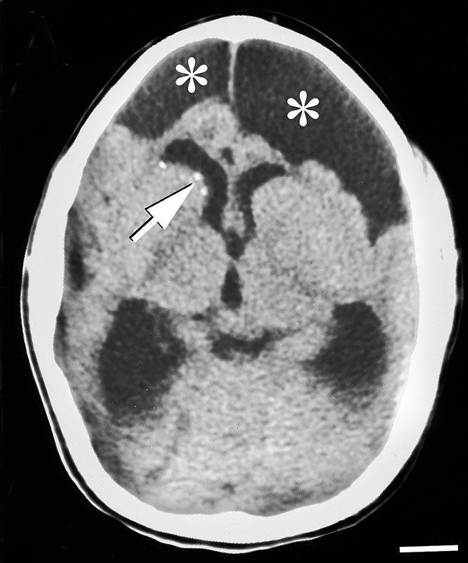 | 665 × 800 (69 KB) | Z8600021 (talk | contribs) | ==Congenital Lymphocytic choriomeningitis virus LCMV infection Computed Tomography== Head CT scan from a 4 month old child with congenital LCMV infection. The scan reveals bilateral asymmetric regions of encephalomalacia (asterisks), strongly suggestiv |
You cannot overwrite this file.
File usage
The following page uses this file: