File:Corpus luteum.jpg: Difference between revisions
(uploaded a new version of "File:Corpus luteum.jpg") |
No edit summary |
||
| Line 1: | Line 1: | ||
==Ovary - Corpus Luteum== | |||
Histology image shows the theca lutein cells and granulosa lutein cells. These cells work together in the production of for | |||
===Theca Lutein Cells=== | |||
* the darker stained cells. | |||
* derived from the theca interna of the original follicle. | |||
* lack microvilli on the surface. | |||
* lack the aromatase enzyme. | |||
* produce androgens for the granulosa lutein cells to convert. | |||
[[Category: | ===Granulosa Lutein Cells=== | ||
* the lighter stained cells. | |||
* derived from the granulosa cells of the original follicle. | |||
* contain aromatase enzyme. | |||
* produce estrogen and progesterone from the androgens produced by the theca lutein cells. | |||
:'''Links:''' [[:File:Corpus luteum.jpg|corpus luteum - low power]] | [[:File:Corpus_luteum_lutein_cells.jpgcorpus luteum - high power]] | |||
{{Blue Histology}} | |||
Histology image H&E high power Clu01he.jpg | |||
[[Category:Histology]] [[Category:Genital]] [[Category:Ovary]] | |||
Revision as of 17:01, 6 May 2012
Ovary - Corpus Luteum
Histology image shows the theca lutein cells and granulosa lutein cells. These cells work together in the production of for
Theca Lutein Cells
- the darker stained cells.
- derived from the theca interna of the original follicle.
- lack microvilli on the surface.
- lack the aromatase enzyme.
- produce androgens for the granulosa lutein cells to convert.
Granulosa Lutein Cells
- the lighter stained cells.
- derived from the granulosa cells of the original follicle.
- contain aromatase enzyme.
- produce estrogen and progesterone from the androgens produced by the theca lutein cells.
Links: Histology | Histology Stains | Blue Histology images copyright Lutz Slomianka 1998-2009. The literary and artistic works on the original Blue Histology website may be reproduced, adapted, published and distributed for non-commercial purposes. See also the page Histology Stains.
Cite this page: Hill, M.A. (2024, June 5) Embryology Corpus luteum.jpg. Retrieved from https://embryology.med.unsw.edu.au/embryology/index.php/File:Corpus_luteum.jpg
- © Dr Mark Hill 2024, UNSW Embryology ISBN: 978 0 7334 2609 4 - UNSW CRICOS Provider Code No. 00098G
Histology image H&E high power Clu01he.jpg
File history
Click on a date/time to view the file as it appeared at that time.
| Date/Time | Thumbnail | Dimensions | User | Comment | |
|---|---|---|---|---|---|
| current | 16:58, 6 May 2012 | 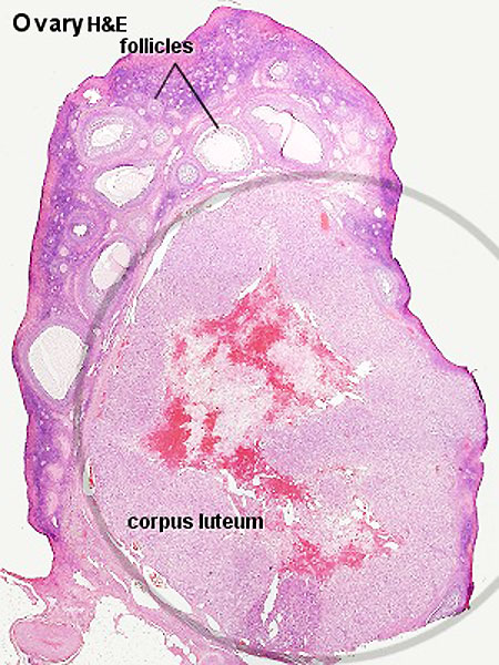 | 450 × 600 (94 KB) | Z8600021 (talk | contribs) | |
| 10:14, 3 August 2009 | 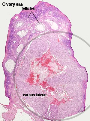 | 300 × 400 (55 KB) | MarkHill (talk | contribs) | Corpus luteum Histology image H&E low power Image Source: Lutz Slomianka, UWA Blue Histology Clu01he.jpg http://www.lab.anhb.uwa.edu.au/mb140/CorePages/FemaleRepro/femalerepro.htm#Corpus |
You cannot overwrite this file.
File usage
The following 11 pages use this file:
- 2010 BGD Practical 3 - Implantation
- 2011 Lab 2 - Week 2
- ANAT2241 Female Reproductive System
- ANAT2341 Lab 2 - Week 2
- BGDA Practical - Female Reproductive Tract Histology
- BGDA Practical 3 - Implantation
- Corpus Luteum Development
- Endocrine System Development
- Human Chorionic Gonadotropin
- Ovary Development
- Talk:2011 Lab 2 - Week 2