File:Eye and retina cartoon.jpg: Difference between revisions
| Line 36: | Line 36: | ||
{{JCB}} | |||
'''Retinal Cell Organization''' and '''Human Retina Histology''' were flipped vertically from original image to match the orientation in '''The Eye'''. This is the ongoing issue of which way around the retina should be illustrated. For an introduction to this topic it is just easier to have it the same way around. | '''Retinal Cell Organization''' and '''Human Retina Histology''' were flipped vertically from original image to match the orientation in '''The Eye'''. This is the ongoing issue of which way around the retina should be illustrated. For an introduction to this topic it is just easier to have it the same way around. | ||
Revision as of 07:42, 5 March 2012
Eye and Retina
The Eye
|
Retinal Cell Organization
|
Human Retina Histology
|
Retina histology image - Swaroop and Zack (2002), published by BioMed Central PMID 12186651
Reference
<pubmed>20855501</pubmed>| PMC3101587 | JCB
Copyright
Rockefeller University Press - Copyright Policy This article is distributed under the terms of an Attribution–Noncommercial–Share Alike–No Mirror Sites license for the first six months after the publication date (see http://www.jcb.org/misc/terms.shtml). After six months it is available under a Creative Commons License (Attribution–Noncommercial–Share Alike 4.0 Unported license, as described at https://creativecommons.org/licenses/by-nc-sa/4.0/ ). (More? Help:Copyright Tutorial)
Retinal Cell Organization and Human Retina Histology were flipped vertically from original image to match the orientation in The Eye. This is the ongoing issue of which way around the retina should be illustrated. For an introduction to this topic it is just easier to have it the same way around.
File history
Click on a date/time to view the file as it appeared at that time.
| Date/Time | Thumbnail | Dimensions | User | Comment | |
|---|---|---|---|---|---|
| current | 14:50, 4 March 2012 | 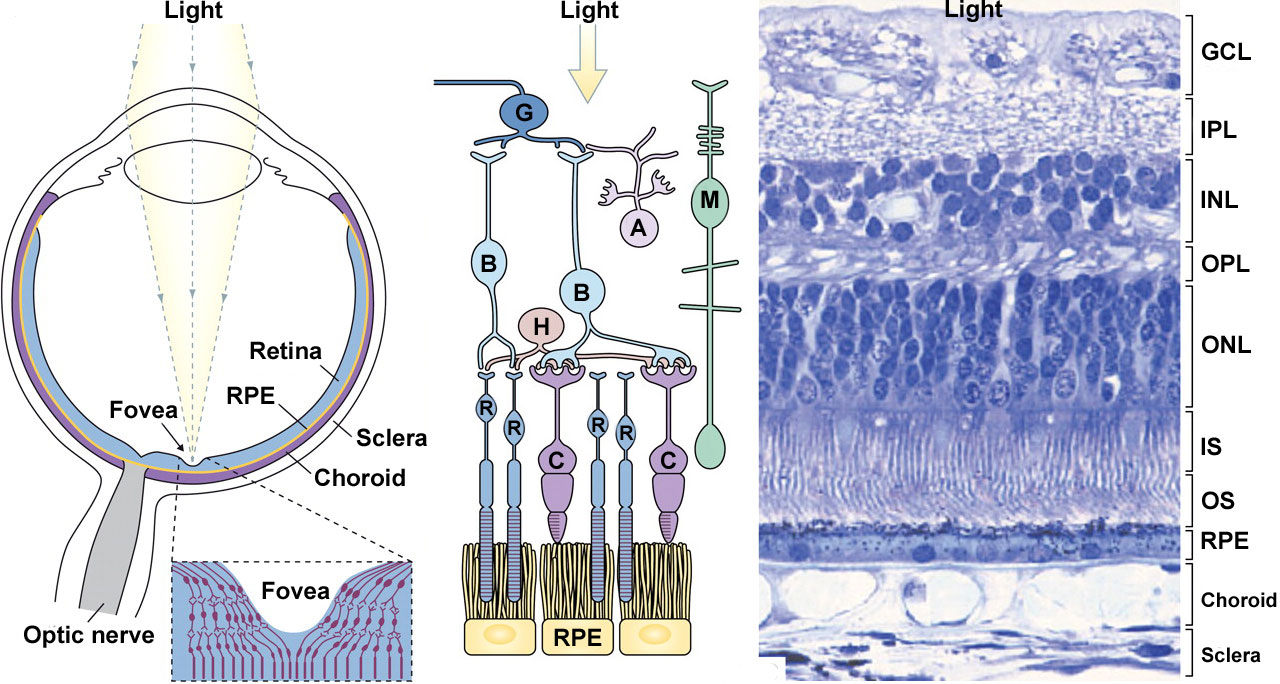 | 1,280 × 684 (211 KB) | Z8600021 (talk | contribs) |
You cannot overwrite this file.