File:Eye and retina cartoon.jpg: Difference between revisions
From Embryology
| Line 2: | Line 2: | ||
{| | {| | ||
| valign="top"|'''The Eye''' | | valign="top"|width="150px"|'''The Eye''' | ||
* An enlarged diagram of the fovea is shown in the box. | * An enlarged diagram of the fovea is shown in the box. | ||
| Line 8: | Line 8: | ||
* The RPE is sandwiched between the retina and choroids, a vascularized and pigmented connective tissue. | * The RPE is sandwiched between the retina and choroids, a vascularized and pigmented connective tissue. | ||
| valign="top"|'''Retinal Cell Organization''' | | valign="top"|width="150px"|'''Retinal Cell Organization''' | ||
* '''G''' - ganglion cells | * '''G''' - ganglion cells | ||
* '''M''' - Müller cell | * '''M''' - Müller cell | ||
Revision as of 14:57, 4 March 2012
Eye and Retina
width="150px"|The Eye
|
width="150px"|Retinal Cell Organization
|
Human Retina Histology
|
Retina histology image - Swaroop and Zack (2002), published by BioMed Central.
File history
Click on a date/time to view the file as it appeared at that time.
| Date/Time | Thumbnail | Dimensions | User | Comment | |
|---|---|---|---|---|---|
| current | 14:50, 4 March 2012 | 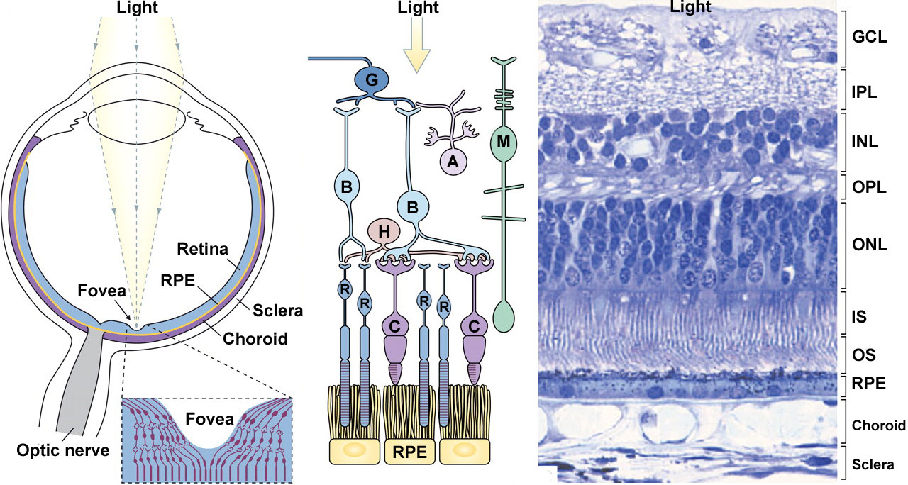 | 1,280 × 684 (211 KB) | Z8600021 (talk | contribs) |
You cannot overwrite this file.