File:Lymphocyte rosettes EM01-06.jpg: Difference between revisions
(Sheep Red Blood Cells Fig. 1. SRBC washed with HBSS showing spheroechinocyte transformation. Two SRBC have maintained their biconcaveshape. X 3,600. Fig. 2. SRBC showing microspherulation, with multiple microprojections with beaded ends. X 6,900. Fig. ) |
No edit summary |
||
| Line 26: | Line 26: | ||
{{JCB}} | {{JCB}} | ||
[[Category:Immune]] [[Category:Electron Micrograph]] [[Category:Scanning EM]] | |||
Revision as of 12:48, 22 February 2012
Sheep Red Blood Cells
Fig. 1. SRBC washed with HBSS showing spheroechinocyte transformation. Two SRBC have maintained their biconcaveshape. X 3,600.
Fig. 2. SRBC showing microspherulation, with multiple microprojections with beaded ends. X 6,900.
Fig. 3. E-rosette showing a smooth surfaced T lymphocyte surrounded by SRBC; the majority of T cells and less than 10% of rosetting B ceils had this type of surface. X 5,700. FIO.
Fig. 4. E-rosette showing intermediate type of T lymphocyte.Up to 30% of rosetting B and T cells had this type of surface. X 6,000.
Fig. 5. EAC-rosette, showing villous B cell. The majority of B cells and about 15% of T cells showed this surface morphology.X 5,700.
Fig. 6. Rosetting T lymphocyte with a villous surface, surrounded by spheroechinocyte SRBC which have multiple projections with beaded extremities of similar dimensions to lymphocyte microvilli. This type of picture suggested that deposition of portions of SRBC projections to the lymphocyte surface may occur. X 6,600.
Lymphocyte rosettes EM01-06.jpg
References
4545895
http://www.ncbi.nlm.nih.gov/pmc/articles/PMC2139697
Copyright
Rockefeller University Press - Copyright Policy This article is distributed under the terms of an Attribution–Noncommercial–Share Alike–No Mirror Sites license for the first six months after the publication date (see http://www.jcb.org/misc/terms.shtml). After six months it is available under a Creative Commons License (Attribution–Noncommercial–Share Alike 4.0 Unported license, as described at https://creativecommons.org/licenses/by-nc-sa/4.0/ ). (More? Help:Copyright Tutorial)
File history
Click on a date/time to view the file as it appeared at that time.
| Date/Time | Thumbnail | Dimensions | User | Comment | |
|---|---|---|---|---|---|
| current | 12:47, 22 February 2012 | 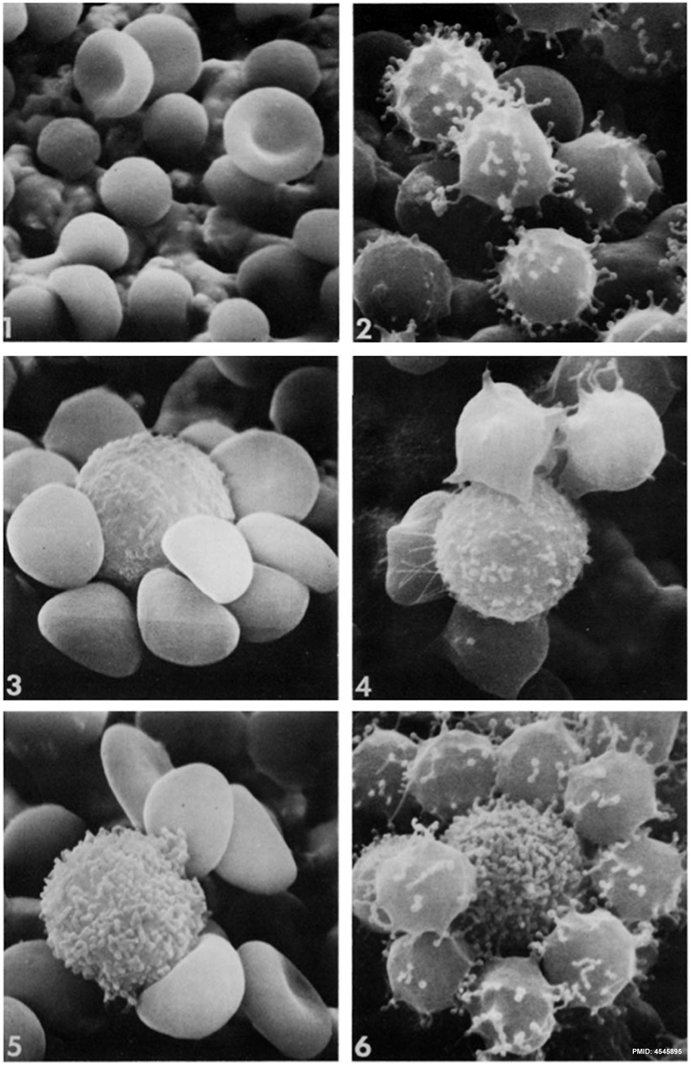 | 1,364 × 2,100 (334 KB) | Z8600021 (talk | contribs) | Sheep Red Blood Cells Fig. 1. SRBC washed with HBSS showing spheroechinocyte transformation. Two SRBC have maintained their biconcaveshape. X 3,600. Fig. 2. SRBC showing microspherulation, with multiple microprojections with beaded ends. X 6,900. Fig. |
You cannot overwrite this file.
File usage
The following 3 pages use this file: