Category:Notochord: Difference between revisions
From Embryology
(Created page with 'The notochord (axial mesoderm) is a rod of cells lying in the midline of the trilaminar embryo mesoderm layer ventral to the neural tube. Thought to have at least 2 early roles i…') |
No edit summary |
||
| Line 2: | Line 2: | ||
The pages and images below relate to the notochord. | The pages and images below relate to the notochord. | ||
[[Category:Week 3]] | |||
Revision as of 07:18, 22 November 2011
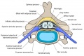
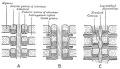
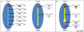
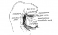
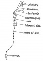
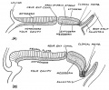
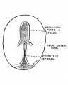
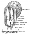
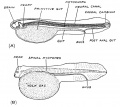
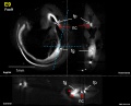
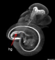
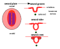
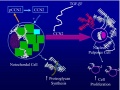
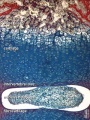
The notochord (axial mesoderm) is a rod of cells lying in the midline of the trilaminar embryo mesoderm layer ventral to the neural tube. Thought to have at least 2 early roles in development and later roles in patterning surrounding tissues. 1. Mechanical, influencing the folding of the early embryo; 2. Morphogenic, secreting sonic hedgehog a protein which regulates the development of surrounding tissues (neural plate, somites, endoderm and other organs). In humans, the notochord forms in week 3 and is eventually lost during the formation of the vertebral column.
The pages and images below relate to the notochord.
Pages in category 'Notochord'
The following 23 pages are in this category, out of 23 total.
P
R
Media in category 'Notochord'
The following 32 files are in this category, out of 32 total.
 Cervical vertebra.jpg 767 × 514; 71 KB
 Gray0065.jpg 600 × 345; 39 KB
 Human notochord development theories 1.jpg 1,280 × 496; 64 KB
 Keith1902 fig015a.jpg 971 × 600; 74 KB
 Keith1902 fig117.jpg 561 × 800; 46 KB
 Keith1921 fig035.jpg 984 × 808; 143 KB
 Keith1921 fig036.jpg 547 × 686; 42 KB
 Keith1921 fig037.jpg 612 × 687; 102 KB
 Keith1921 fig040.jpg 901 × 806; 138 KB
 Kollmann057.jpg 845 × 469; 49 KB
 Mouse E11 Foxf1.jpg 1,420 × 1,143; 162 KB
 Mouse E11 Sox2 and Nkx2.1.jpg 2,229 × 720; 176 KB
 Mouse E9 Foxf1.jpg 1,420 × 1,143; 142 KB
 Mouse embryo E11 HNF3beta notochord marker 01.jpg 2,245 × 829; 129 KB
 Mouse embryo E11 HNF3beta notochord marker 02.jpg 913 × 1,000; 68 KB
 Mouse embryo E11 HNF3beta notochord marker 03.jpg 913 × 1,000; 56 KB
 Mouse embryo E11 HNF3beta notochord marker 04.jpg 913 × 1,000; 55 KB
 Neuralplate cartoon.png 343 × 284; 5 KB
 Notochordal interaction with nucleus pulposus.jpg 478 × 360; 92 KB
 Ossification endochondral 01.jpg 817 × 613; 198 KB
 Ossification endochondral 1.jpg 750 × 1,000; 147 KB
 Ossification endochondral 1c.jpg 300 × 400; 32 KB
 Shh frog notochord 1.jpg 150 × 150; 8 KB
 Stage 13 image 056.jpg 1,000 × 516; 102 KB
 Stage 13 image 057.jpg 1,000 × 511; 99 KB
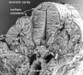 Stage11 sem100.jpg 1,000 × 898; 109 KB
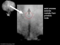 Stage7 axial process.jpg 500 × 375; 11 KB
 Williams1908-fig17.jpg 982 × 1,500; 145 KB
 Williams1908-fig18.jpg 921 × 1,500; 89 KB
 Williams1908-fig19.jpg 981 × 1,500; 104 KB
 Williams1908-fig20.jpg 920 × 1,500; 96 KB
 Xenopus FoxA4 model.jpg 1,192 × 565; 104 KB


