File:Fetal head lateral.jpg: Difference between revisions
From Embryology
(bone and cartilage Headbone.jpg) |
No edit summary |
||
| Line 1: | Line 1: | ||
bone and cartilage | Fetal Head (12 weeks) | ||
Headbone.jpg | |||
lateral view, stained for bone and cartilage | |||
In this lateral (external) view, note the distribution of new bone in the plates of the cranial vault, temporal bone, orbit, upper jaw (maxilla) and lower jaw (mandible) regions. (More? Notes Skull | Face) Bony regions in the lower jaw (mandible) region also show spaces where tooth formation is occuring. (More? Tooth Development) | |||
Original File Name: Headbone.jpg | |||
Source: UNSW Embryology http://embryology.med.unsw.edu.au/wwwhuman/Hum12wk/Hum12wk.htm | |||
Revision as of 15:48, 13 September 2009
Fetal Head (12 weeks)
lateral view, stained for bone and cartilage
In this lateral (external) view, note the distribution of new bone in the plates of the cranial vault, temporal bone, orbit, upper jaw (maxilla) and lower jaw (mandible) regions. (More? Notes Skull | Face) Bony regions in the lower jaw (mandible) region also show spaces where tooth formation is occuring. (More? Tooth Development)
Original File Name: Headbone.jpg
Source: UNSW Embryology http://embryology.med.unsw.edu.au/wwwhuman/Hum12wk/Hum12wk.htm
File history
Click on a date/time to view the file as it appeared at that time.
| Date/Time | Thumbnail | Dimensions | User | Comment | |
|---|---|---|---|---|---|
| current | 15:44, 13 September 2009 | 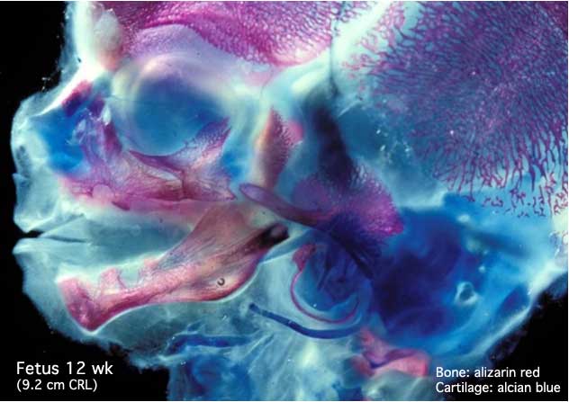 | 632 × 447 (34 KB) | S8600021 (talk | contribs) | bone and cartilage Headbone.jpg |
You cannot overwrite this file.
File usage
The following 52 pages use this file:
- 2009 Lecture 13
- 2009 Lecture 18
- 2010 BGD Practical 12 - Second Trimester
- 2010 Lab 6
- 2010 Lecture 11
- 2010 Lecture 13
- 2010 Lecture 22
- 2011 Lab 12 - Second Trimester
- 2011 Lab 6 - Fetal
- 2011 Lab 6 - Postnatal
- 2011 Lab 9
- AACP Meeting 2013 - Face Embryology
- ANAT2241 Bone, Bone Formation and Joints
- ANAT2341 Lab 11 - Embryo to Fetus
- ANAT2341 Lab 11 - Second Trimester
- ANAT2341 Lab 12 - Second Trimester
- ANAT2341 Lab 6 - Fetal
- ANAT2341 Lab 6 - Postnatal
- Abnormal Development - Fetal Growth Restriction
- BGDA Practical 12 - Second Trimester
- BGDB Face and Ear - Fetal
- BGD Lecture - Face and Ear Development
- Bone Development
- Bone Histology
- Cartilage Histology
- Fetal Development
- Fetal Development - 12 Weeks
- Head Development
- Joint Development - Temporomandibular Joint
- Lecture - Head Development
- Lecture - Musculoskeletal Development
- M
- Musculoskeletal System - Abnormalities
- Musculoskeletal System - Axial Skeleton Development
- Musculoskeletal System - Bone Development
- Musculoskeletal System - Bone Development Timeline
- Musculoskeletal System - Cartilage Development
- Musculoskeletal System - Joint Development
- Musculoskeletal System - Skull Development
- Musculoskeletal System Development
- Second Trimester
- Timeline human development
- Virginia Diewert
- Talk:ANAT2341 Lab 7
- Talk:ANAT2341 Lab 9
- Talk:Timeline human development
- Template:Second Trimester Timeline
- Template:Second Trimester Timeline collapsable table
- Template:Second Trimester table01
- Template talk:Second Trimester Timeline
- Category:Fetal
- Category:Second Trimester