File:Stage 22 image 221.jpg: Difference between revisions
From Embryology
| Line 10: | Line 10: | ||
:'''Links:''' [[:File:Stage_22_image_220.jpg|Image - Pituitary]] | [[:File:Stage_22_image_221.jpg|Image - Pituitary Measured]] | [[Endocrine - Pituitary Development]] | [[:File:Stage_22_image_156.jpg|Image - Low resolution Plane of Section]] | [[:File:Stage_22_image_158.jpg|Image - Low resolution Pituitary]] | :'''Links:''' [[:File:Stage_22_image_220.jpg|Image - Pituitary]] | [[:File:Stage_22_image_221.jpg|Image - Pituitary Measured]] | [[Endocrine - Pituitary Development]] | [[Carnegie stage 22]] | [[:File:Stage_22_image_156.jpg|Image - Low resolution Plane of Section]] | [[:File:Stage_22_image_158.jpg|Image - Low resolution Pituitary]] | ||
Revision as of 07:12, 29 August 2011
Human Embryo Pituitary
- Week 8, Carnegie Stage 22
- inset bottom right shows the complete head cross-section and approximate level of the plane.
- note the sphenoid "saddle"
- trigeminal ganglia (CN V) and internal carotid arteries lateral to pituitary in complete head cross-section.
- orientation - ventral is bottom of image, dorsal is top.
- inset cross-section scale bar 1 mm.
- measured regions are shown in microns.
- Links: Image - Pituitary | Image - Pituitary Measured | Endocrine - Pituitary Development | Carnegie stage 22 | Image - Low resolution Plane of Section | Image - Low resolution Pituitary
A7/B2 - Scan 35805
File history
Click on a date/time to view the file as it appeared at that time.
| Date/Time | Thumbnail | Dimensions | User | Comment | |
|---|---|---|---|---|---|
| current | 06:49, 29 August 2011 | 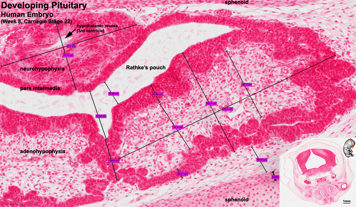 | 1,200 × 699 (336 KB) | S8600021 (talk | contribs) |
You cannot overwrite this file.
File usage
The following 37 pages use this file:
- 2011 Lab 9
- BGDA Practical 7 - Week 8
- Carnegie stage 22
- Endocrine - Pituitary Development
- Talk:ANAT2341 Lab 7
- Talk:ANAT2341 Lab 9
- File:Stage22 vertebra and spinal cord 1.jpg
- File:Stage 22 image 200.jpg
- File:Stage 22 image 201.jpg
- File:Stage 22 image 203.jpg
- File:Stage 22 image 204.jpg
- File:Stage 22 image 205.jpg
- File:Stage 22 image 206.jpg
- File:Stage 22 image 207.jpg
- File:Stage 22 image 208.jpg
- File:Stage 22 image 209.jpg
- File:Stage 22 image 210.jpg
- File:Stage 22 image 211.jpg
- File:Stage 22 image 212.jpg
- File:Stage 22 image 213.jpg
- File:Stage 22 image 214.jpg
- File:Stage 22 image 215.jpg
- File:Stage 22 image 216.jpg
- File:Stage 22 image 217.jpg
- File:Stage 22 image 218.jpg
- File:Stage 22 image 219.jpg
- File:Stage 22 image 220.jpg
- File:Stage 22 image 222.jpg
- File:Stage 22 image 223.jpg
- File:Stage 22 image 224.jpg
- File:Stage 22 image 225.jpg
- File:Stage 22 image 301.jpg
- File:Stage 22 image 302.jpg
- File:Stage 22 image 322.jpg
- File:Stage 22 vomeronasal organ.jpg
- Template:Stage 22 histology gallery
- Template:Stage 22 histology gallery table