File:Pancreas histology 102.jpg: Difference between revisions
From Embryology
No edit summary |
No edit summary |
||
| Line 1: | Line 1: | ||
==Pancreas== | ==Pancreas== | ||
* The exocrine pancreas consists of tubuloacinar glands. | |||
* A single layer of pyramidal shaped cells forms the secretory acini. | |||
* The apical cytoplasm (towards the lumen of the acini) is filled with secretory vesicles containing the precursors of digestive enzymes. | |||
* The first portion of the duct system extends into the centre of the acini, which is lined by small centroacinar cells. | |||
* These cells form the first part of intercalated ducts. | |||
* Intercalated ducts are lined by low columnar or cuboidal epithelium. | |||
* They empty into interlobular ducts, which are lined by a columnar epithelium. | |||
* Interlobular ducts in turn empty into the main pancreatic duct (of Wirsung), which is lined by a tall columnar epithelium. | |||
{{Pancreas Histology Images}} | {{Pancreas Histology Images}} | ||
Latest revision as of 12:23, 9 August 2011
Pancreas
- The exocrine pancreas consists of tubuloacinar glands.
- A single layer of pyramidal shaped cells forms the secretory acini.
- The apical cytoplasm (towards the lumen of the acini) is filled with secretory vesicles containing the precursors of digestive enzymes.
- The first portion of the duct system extends into the centre of the acini, which is lined by small centroacinar cells.
- These cells form the first part of intercalated ducts.
- Intercalated ducts are lined by low columnar or cuboidal epithelium.
- They empty into interlobular ducts, which are lined by a columnar epithelium.
- Interlobular ducts in turn empty into the main pancreatic duct (of Wirsung), which is lined by a tall columnar epithelium.
- Pancreas Histology Links: overview (label) | exocrine (label) | endocrine (label) | blood vessels (label) | insulin (label) | overview | exocrine | endocrine | blood vessels | insulin | Islet labeled for insulin and Glucagon | Insulin (Fl) | Glucagon (Fl) | GIT Histology
Links: Histology | Histology Stains | Blue Histology images copyright Lutz Slomianka 1998-2009. The literary and artistic works on the original Blue Histology website may be reproduced, adapted, published and distributed for non-commercial purposes. See also the page Histology Stains.
Cite this page: Hill, M.A. (2024, June 14) Embryology Pancreas histology 102.jpg. Retrieved from https://embryology.med.unsw.edu.au/embryology/index.php/File:Pancreas_histology_102.jpg
- © Dr Mark Hill 2024, UNSW Embryology ISBN: 978 0 7334 2609 4 - UNSW CRICOS Provider Code No. 00098G
File history
Click on a date/time to view the file as it appeared at that time.
| Date/Time | Thumbnail | Dimensions | User | Comment | |
|---|---|---|---|---|---|
| current | 23:21, 9 July 2011 | 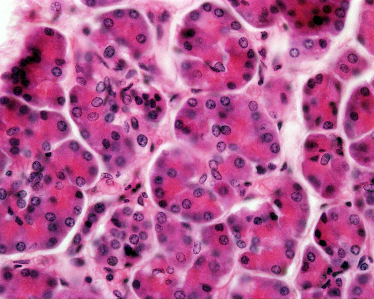 | 1,280 × 1,024 (309 KB) | S8600021 (talk | contribs) |
You cannot overwrite this file.
File usage
The following 3 pages use this file: