File:Stage14 sem2l.jpg: Difference between revisions
No edit summary |
No edit summary |
||
| Line 12: | Line 12: | ||
{{Template:Footer}} | {{Template:Footer}} | ||
[[Category:Carnegie | [[Category:Carnegie Stage 14]] [[Category:Placode]] [[Category:Head]] | ||
Revision as of 14:48, 9 June 2011
Human Embryo Carnegie stage 14
Carnegie stage 14, day 32, 35 somites
Original file name: Stage14day32somite35-ventral-sem2-1000px.jpg
Image Code: Stage14= carnegie stage sem= scanning em 2 = image 2 1000px size l=labeled
Image Source: Scanning electron micrographs of the Carnegie stages of the early human embryos are reproduced with the permission of Prof Kathy Sulik, from embryos collected by Dr. Vekemans and Tania Attié-Bitach. Images are for educational purposes only and cannot be reproduced electronically or in writing without permission.
Cite this page: Hill, M.A. (2024, June 2) Embryology Stage14 sem2l.jpg. Retrieved from https://embryology.med.unsw.edu.au/embryology/index.php/File:Stage14_sem2l.jpg
- © Dr Mark Hill 2024, UNSW Embryology ISBN: 978 0 7334 2609 4 - UNSW CRICOS Provider Code No. 00098G
File history
Click on a date/time to view the file as it appeared at that time.
| Date/Time | Thumbnail | Dimensions | User | Comment | |
|---|---|---|---|---|---|
| current | 10:16, 3 September 2009 | 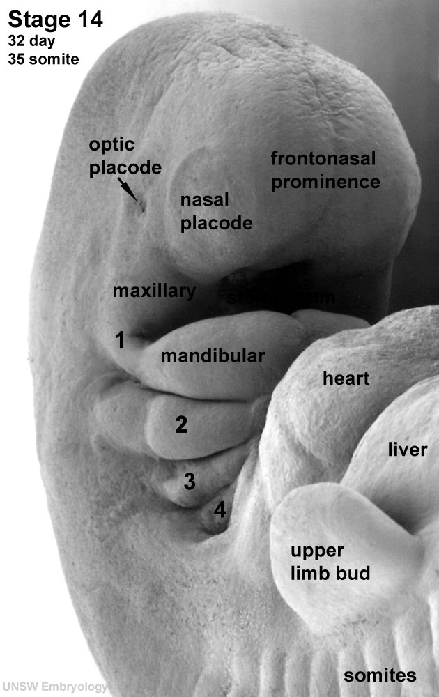 | 630 × 1,000 (96 KB) | S8600021 (talk | contribs) | Human Embryo Carnegie stage 14, day 32, 35 somites Original file name: Stage14day32somite35-ventral-sem2-1000px.jpg Image Code: Stage14= carnegie stage sem= scanning em 2 = image 2 1000px size l=labeled {{Template:SEM}} {{Template:Footer}} |
You cannot overwrite this file.
File usage
The following 18 pages use this file:
- 2011 Lab 10 - Early Embryo
- 2011 Lab 6 - Early Embryo
- AACP Meeting 2013 - Face Embryology
- ANAT2341 Lab 10 - Early Embryo
- ANAT2341 Lab 3 - Week 4
- Abnormal Development - Thalidomide
- B
- BGDA Lecture - Development of the Embryo/Fetus 2
- Human Embryo SEM
- K12 Comparative Embryology
- K12 Thalidomide
- Lecture - Head Development
- Lecture - Sensory Development
- Musculoskeletal System - Appendicular Skeleton Development
- Musculoskeletal System - Limb Development
- Pharyngeal arches
- Sensory System Development
- Template:Carnegie stage 11-14 image table