File:Spina bifida.jpg: Difference between revisions
No edit summary |
|||
| Line 17: | Line 17: | ||
This is an Open Access article distributed under the terms of the Creative Commons Attribution License (http://creativecommons.org/licenses/by/2.0), which permits unrestricted use, distribution, and reproduction in any medium, provided the original work is properly cited. | This is an Open Access article distributed under the terms of the Creative Commons Attribution License (http://creativecommons.org/licenses/by/2.0), which permits unrestricted use, distribution, and reproduction in any medium, provided the original work is properly cited. | ||
[[Category:Human]] [[Category:Abnormal Development]] [[Category:Musculoskeletal]] [[Category:Bone]] [[Category: | [[Category:Human]] [[Category:Abnormal Development]] [[Category:Musculoskeletal]] [[Category:Bone]] [[Category:Computed Tomography]] | ||
Revision as of 23:56, 2 May 2011
Spina Bifida
Preoperative three dimensional CT scan. A-P view (A) and lateral view (B) demonstrating severe kyphosis at the lumbar region and spina bifida below L1.
A 32-year-old woman was referred to our hospital for a refractory ulcer on her back. She had a history of myelomeningocele with spina bifida that was treated surgically at birth.
Original file name: Figure 3 1748-7161-6-5-3.jpg http://www.ncbi.nlm.nih.gov/pmc/articles/PMC3080349/figure/F3/
Reference
<pubmed>21477271</pubmed>| PMC3080349 | Scoliosis.
Scoliosis. 2011; 6: 5.
Published online 2011 April 8. doi: 10.1186/1748-7161-6-5.
This is an Open Access article distributed under the terms of the Creative Commons Attribution License (http://creativecommons.org/licenses/by/2.0), which permits unrestricted use, distribution, and reproduction in any medium, provided the original work is properly cited.
File history
Click on a date/time to view the file as it appeared at that time.
| Date/Time | Thumbnail | Dimensions | User | Comment | |
|---|---|---|---|---|---|
| current | 23:50, 2 May 2011 | 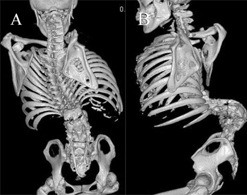 | 800 × 633 (77 KB) | S8600021 (talk | contribs) | ==Spina Bifida== Preoperative three dimensional CT scan. A-P view (A) and lateral view (B) demonstrating severe kyphosis at the lumbar region and spina bifida below L1. Original file name: Figure 3 1748-7161-6-5-3.jpg http://www.ncbi.nlm.nih.gov/pmc/ar |
You cannot overwrite this file.
File usage
There are no pages that use this file.