File:Human holoprosencephaly cyclopia dissection.jpg: Difference between revisions
(Photographs of the macroscopic appearance of the head. a) Frontal view of the investigated head. b) View of the opened cranium with remnants of brain. Link: http://www.pubmedcentral.nih.gov/articlerender.fcgi?artid=2709107&rendertype=figure&id=F1 Ori) |
mNo edit summary |
||
| (9 intermediate revisions by 2 users not shown) | |||
| Line 1: | Line 1: | ||
==Holoprosencephaly and Cyclopia== | |||
Photographs of the macroscopic appearance of the fetal head. | |||
'''a)''' Frontal view of the investigated head. | |||
'''b)''' View of the opened cranium with remnants of brain. | |||
[[International Classification of Diseases]] - Q04 Other congenital malformations of brain - Q04.2 Holoprosencephaly | |||
:[[Neural_System_-_Abnormalities|'''Neural Abnormality Links''']]: [[:File:Human holoprosencephaly cyclopia dissection.jpg|Human holoprosencephaly cyclopia dissection]] | [[:File:Proboscis histology.jpg|Proboscis histology]] | [[Neural_System_-_Abnormalities#Holoprosencephaly|Holoprosencephaly]] | [[Neural_System_-_Abnormalities|Neural Abnormalities]] | [[Developmental Signals - Sonic hedgehog|Sonic hedgehog]] | |||
===Reference=== | |||
{{#pmid:19563629}} | |||
====Copyright==== | |||
Copyright © 2009 Arnold and Meiselbach; licensee BioMed Central Ltd. | |||
This is an Open Access article distributed under the terms of the Creative Commons Attribution License (http://creativecommons.org/licenses/by/2.0), which permits unrestricted use, distribution, and reproduction in any medium, provided the original work is properly cited. | This is an Open Access article distributed under the terms of the Creative Commons Attribution License (http://creativecommons.org/licenses/by/2.0), which permits unrestricted use, distribution, and reproduction in any medium, provided the original work is properly cited. | ||
| Line 16: | Line 27: | ||
Published online 2009 June 29. doi: 10.1186/1746-160X-5-14. | Published online 2009 June 29. doi: 10.1186/1746-160X-5-14. | ||
PMCID: PMC2709107 | PMCID: PMC2709107 | ||
Link: http://www.pubmedcentral.nih.gov/articlerender.fcgi?artid=2709107&rendertype=figure&id=F1 | |||
Original File name: 1746-160X-5-14-1.jpg | |||
{{Footer}} | |||
[[Category:Human]] [[Category:Abnormal Development]] [[Category:Neural]] [[Category:Head]] | |||
Latest revision as of 13:30, 18 July 2019
Holoprosencephaly and Cyclopia
Photographs of the macroscopic appearance of the fetal head.
a) Frontal view of the investigated head.
b) View of the opened cranium with remnants of brain.
International Classification of Diseases - Q04 Other congenital malformations of brain - Q04.2 Holoprosencephaly
- Neural Abnormality Links: Human holoprosencephaly cyclopia dissection | Proboscis histology | Holoprosencephaly | Neural Abnormalities | Sonic hedgehog
Reference
Arnold WH & Meiselbach V. (2009). 3-D reconstruction of a human fetus with combined holoprosencephaly and cyclopia. Head Face Med , 5, 14. PMID: 19563629 DOI.
Copyright
Copyright © 2009 Arnold and Meiselbach; licensee BioMed Central Ltd.
This is an Open Access article distributed under the terms of the Creative Commons Attribution License (http://creativecommons.org/licenses/by/2.0), which permits unrestricted use, distribution, and reproduction in any medium, provided the original work is properly cited.
Head Face Med. 2009; 5: 14. Published online 2009 June 29. doi: 10.1186/1746-160X-5-14. PMCID: PMC2709107
Link: http://www.pubmedcentral.nih.gov/articlerender.fcgi?artid=2709107&rendertype=figure&id=F1 Original File name: 1746-160X-5-14-1.jpg
Cite this page: Hill, M.A. (2024, June 1) Embryology Human holoprosencephaly cyclopia dissection.jpg. Retrieved from https://embryology.med.unsw.edu.au/embryology/index.php/File:Human_holoprosencephaly_cyclopia_dissection.jpg
- © Dr Mark Hill 2024, UNSW Embryology ISBN: 978 0 7334 2609 4 - UNSW CRICOS Provider Code No. 00098G
File history
Click on a date/time to view the file as it appeared at that time.
| Date/Time | Thumbnail | Dimensions | User | Comment | |
|---|---|---|---|---|---|
| current | 08:44, 9 August 2009 | 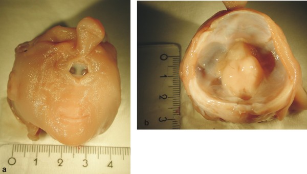 | 600 × 340 (37 KB) | S8600021 (talk | contribs) | Photographs of the macroscopic appearance of the head. a) Frontal view of the investigated head. b) View of the opened cranium with remnants of brain. Link: http://www.pubmedcentral.nih.gov/articlerender.fcgi?artid=2709107&rendertype=figure&id=F1 Ori |
You cannot overwrite this file.
File usage
The following 2 pages use this file: