File:Stage11 sem4.jpg: Difference between revisions
No edit summary |
mNo edit summary |
||
| (18 intermediate revisions by 2 users not shown) | |||
| Line 1: | Line 1: | ||
==Human Embryo Head (Carnegie stage 11)== | |||
{| | |||
| width=400px| | |||
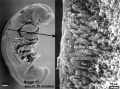
* [[Carnegie stage 11]], [[Week 4]] (25 days, 20 somite pairs), ventral view. | |||
* {{GA}} week 6. | |||
* Ventral view - brain fold, stomodeum, buccopharyngeal membrane, pharyngeal arches, primitive heart. | |||
Carnegie stage 11 | :[[Carnegie stage 11|'''Stage 11 Links''']]: [[:File:Stage11_bf9.jpg|bright field image]] | [[:File:Stage11_sem2.jpg|Detail - buccopharyngeal membrane]] | ||
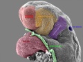
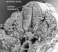
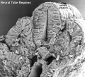
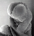
| valign=top|Ventral view of the Week 4 embryo head, showing the stomodeum (stomatodeum or stomatodaeum) region with the floor of this region occupied by the [[:File:Stage11_sem2.jpg|degenerating buccopharyngeal membrane]] (oropharyngeal membrane, stomodeal membrane). Loss of buccopharyngeal membrane opens the gastrointestinal and respiratory tract to amniotic fluid for the remainder of development. | |||
A [[:File:Stage11_bf9.jpg|bright field version]] of this image is also available. | |||
|} | |||
===Relative to the Stomodeum=== | |||
* '''Cranial''' (above) lies brain bulge covered in thin layer of surface ectoderm (surface crack is processing artefact). | |||
* '''Lateral''' (beside) lie the primordia of the first pharyngeal arch. | |||
* '''Inferior''' (below) lies the developing heart (lower left of image), ventral body wall removed to show heart tube. | |||
{{Carnegie stage 11 links}} | |||
{{Carnegie_stage_table_1}} | |||
'''Image version links:''' [[:File:Stage11 sem4.jpg|Large 1000px]] | [[:File:Stage11 sem4a.jpg| 800px]] | | '''Image version links:''' [[:File:Stage11 sem4.jpg|Large 1000px]] | [[:File:Stage11 sem4a.jpg| 800px]] | [[:File:Stage11 sem4b.jpg|Medium 600px]] | ||
[[:File:Stage11 sem4b.jpg|Medium 600px | |||
{{Stage11SEM}} | |||
{{SEM}} | |||
{{ | {{Footer}} | ||
[[Category:Carnegie Stage 11]] | [[Category:Carnegie Stage 11]] | ||
[[Category:Week 4]] | [[Category:Week 4]] | ||
[[Category:Gastrointestinal Tract]] | |||
[[Category:Head]] | |||
Latest revision as of 12:52, 12 May 2019
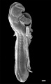
Human Embryo Head (Carnegie stage 11)
|
Ventral view of the Week 4 embryo head, showing the stomodeum (stomatodeum or stomatodaeum) region with the floor of this region occupied by the degenerating buccopharyngeal membrane (oropharyngeal membrane, stomodeal membrane). Loss of buccopharyngeal membrane opens the gastrointestinal and respiratory tract to amniotic fluid for the remainder of development.
A bright field version of this image is also available. |
Relative to the Stomodeum
- Cranial (above) lies brain bulge covered in thin layer of surface ectoderm (surface crack is processing artefact).
- Lateral (beside) lie the primordia of the first pharyngeal arch.
- Inferior (below) lies the developing heart (lower left of image), ventral body wall removed to show heart tube.
| Week: | 1 | 2 | 3 | 4 | 5 | 6 | 7 | 8 |
| Carnegie stage: | 1 2 3 4 | 5 6 | 7 8 9 | 10 11 12 13 | 14 15 | 16 17 | 18 19 | 20 21 22 23 |
Image version links: Large 1000px | 800px | Medium 600px
- Stage 11 SEM Images: dorsolateral whole embryo | dorsal embryo | lateral embryo | lateral head | lateral head with overlay | embryo cross-section | ventrolateral head | ventrolateral head with overlay | ventral head | buccopharyngeal membrane | neural crest | posterior neuropore | anterior neuropore | Carnegie stage 11
- Human Embryo (stage 11)
dorsolateral whole embryo
dorsal embryo
lateral embryo
lateral head
lateral head with overlay

embryo cross-section
embryo cross-section label
neural cross-section label

ventrolateral head
ventrolateral head with overlay

ventral head

buccopharyngeal membrane
neural crest
posterior neuropore
anterior neuropore


Image Source: Scanning electron micrographs of the Carnegie stages of the early human embryos are reproduced with the permission of Prof Kathy Sulik, from embryos collected by Dr. Vekemans and Tania Attié-Bitach. Images are for educational purposes only and cannot be reproduced electronically or in writing without permission.
Cite this page: Hill, M.A. (2024, May 23) Embryology Stage11 sem4.jpg. Retrieved from https://embryology.med.unsw.edu.au/embryology/index.php/File:Stage11_sem4.jpg
- © Dr Mark Hill 2024, UNSW Embryology ISBN: 978 0 7334 2609 4 - UNSW CRICOS Provider Code No. 00098G
File history
Click on a date/time to view the file as it appeared at that time.
| Date/Time | Thumbnail | Dimensions | User | Comment | |
|---|---|---|---|---|---|
| current | 18:09, 7 September 2009 |  | 808 × 1,000 (129 KB) | S8600021 (talk | contribs) | Stage11day25somite20-ventrolateral-oral-membrane-sem4.jpg |
You cannot overwrite this file.
File usage
The following 67 pages use this file:
- AACP Meeting 2013 - Face Embryology
- ANAT2341 Lab 6 - Early Embryo
- Abnormal Development - Thalidomide
- BGDB Face and Ear - Early Embryo
- BGDB Gastrointestinal - Activity 1
- BGD Lecture - Face and Ear Development
- BGD Lecture - Gastrointestinal System Development
- Buccopharyngeal membrane
- Carnegie stage 11
- Draft 2016
- Gastrointestinal Tract - Mouth Development
- Gastrointestinal Tract - Oesophagus Development
- Gastrointestinal Tract Development
- Human Embryo - Scanning electron microscopy
- Human Embryo SEM
- K12 Thalidomide
- Lecture - Gastrointestinal Development
- Lecture - Gastrointestinal Development 2013
- Lecture - Head Development
- Lecture - Respiratory Development
- SH Lecture - Respiratory System Development
- Talk:Carnegie stage 11
- File:Stage11 sem10.jpg
- File:Stage11 sem100.jpg
- File:Stage11 sem100a.jpg
- File:Stage11 sem100b.jpg
- File:Stage11 sem100c.jpg
- File:Stage11 sem101.jpg
- File:Stage11 sem10a.jpg
- File:Stage11 sem10b.jpg
- File:Stage11 sem10c.jpg
- File:Stage11 sem13.jpg
- File:Stage11 sem13a.jpg
- File:Stage11 sem13b.jpg
- File:Stage11 sem13c.jpg
- File:Stage11 sem2.jpg
- File:Stage11 sem21.jpg
- File:Stage11 sem2a.jpg
- File:Stage11 sem2b.jpg
- File:Stage11 sem2c.jpg
- File:Stage11 sem3.jpg
- File:Stage11 sem3a.jpg
- File:Stage11 sem3b.gif
- File:Stage11 sem3b.jpg
- File:Stage11 sem3c.jpg
- File:Stage11 sem4.jpg
- File:Stage11 sem4a.jpg
- File:Stage11 sem4b.jpg
- File:Stage11 sem4c.jpg
- File:Stage11 sem5.jpg
- File:Stage11 sem5a.jpg
- File:Stage11 sem5b.jpg
- File:Stage11 sem5c.jpg
- File:Stage11 sem6.jpg
- File:Stage11 sem7.jpg
- File:Stage11 sem7a.jpg
- File:Stage11 sem7b.jpg
- File:Stage11 sem8.jpg
- File:Stage11 sem81.jpg
- File:Stage11 sem82.jpg
- File:Stage11 sem8a.jpg
- File:Stage11 sem8b.jpg
- File:Stage11 sem9.jpg
- File:Stage11 sem9a.jpg
- File:Stage11 sem9b.jpg
- Template:Carnegie stage 11-14 image table
- Template:Stage11SEM