Category:Carnegie Embryo 75: Difference between revisions
(Created page with "{| class="wikitable" |- ! Serial No. !! Size (mm) !! Grade !! Fixative !! Embedding Medium !! Plane !! Thinness (µm) !! Stain !! Point Score !! Sex !! Year !! Notes |- | 45 |...") |
mNo edit summary |
||
| (5 intermediate revisions by the same user not shown) | |||
| Line 1: | Line 1: | ||
This {{Embryology}} category shows pages and images that relate to the [[Carnegie Collection]] Embryo No. {{CE75}}. This embryo was classified as [[Carnegie stage 23|Stage 23]]. | |||
<br> | |||
{{Carnegie stage 23 links}} | |||
<br> | |||
{| class="wikitable" | {| class="wikitable" | ||
|- | |- | ||
! Serial No. !! Size (mm) !! Grade !! Fixative !! Embedding Medium !! Plane !! Thinness (µm) !! Stain !! Point Score !! Sex !! Year !! Notes | ! Serial No. !! Size (mm) !! Grade !! Fixative !! Embedding Medium !! Plane !! Thinness (µm) !! Stain !! Point Score !! Sex !! Year !! Notes | ||
|- | |- | ||
| | | {{CE75}} || E,30 || Good || Alc. || P || Sag. || 50 || Coch. || 57 || M || 1897 || | ||
|} | |} | ||
===References=== | |||
{{Ref-Streeter1919}} | |||
{{Ref-Kunitomo1920}} | |||
{{Carnegie Collection stage 23 table}} | {{Carnegie Collection stage 23 table}} | ||
===Embryo No. {{CE75}}, 30 mm Crown-Rump Length=== | |||
{{Ref-Kunitomo1920}} | |||
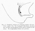
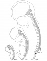
At the caudal end of embryo No. {{CE75}} there is a small papilliform tail containing a group of cells which merge into the wall of the spinal canal, as shown in figure 43. The spinal cord narrows suddenly at the mid-level of the thirty-second vertebra, and its atrophic portion is further constricted at a level between the thirty-third and thirty-fourth vertebrae, as indicated in figure 43 (constrict). The part below this constriction is the primordium of the coccygeal medullary vestige and the upper part is destined in a later stage to undergo retrogression, leaving a small cell-sac as a second coccygeal medullary vestige. There are two large folds on the ventral wall of the spinal cord at a level with the thirty-first vertebra. In the median plane they are triangular in shape and consist of ependymal and mesenchymal cells that have been inverted, together with the wall. A large diverticulum lies between these two folds. The space below the folds probably represents the primordium of the ventriculus terminalis. Only the branches of the anterior spinal artery enter into these folds. There are 34 cartilaginous vertebrae, and at thirty-first and thirty-second vertebrae the column presents a typical curve. The chorda dorsalis shows a spindle-shaped swelling between the vertebrae, and is much convoluted at the caudal end, as seen in figure 43. There are 31 spinal ganglia; the nerves of the last pair are quite slender. | |||
{{Footer}} | |||
[[ | [[Category:Carnegie Stage 23]][[Category:1800's]] | ||
[[Category:Male]] | |||
Latest revision as of 14:40, 25 May 2018
This Embryology category shows pages and images that relate to the Carnegie Collection Embryo No. 75. This embryo was classified as Stage 23.
| Serial No. | Size (mm) | Grade | Fixative | Embedding Medium | Plane | Thinness (µm) | Stain | Point Score | Sex | Year | Notes |
|---|---|---|---|---|---|---|---|---|---|---|---|
| 75 | E,30 | Good | Alc. | P | Sag. | 50 | Coch. | 57 | M | 1897 |
References
Streeter GL. Factors involved in the formation of the filum terminale. (1919) Amer. J Anat. 22(1): 1-11.
Kunitomo K. The development and reduction of the tail and of the caudal end of the spinal cord (1920) Contrib. Embryol., Carnegie Inst. Wash. Publ. 272, 9: 163-198.
| Carnegie Collection - Stage 23 | |||||||||||
|---|---|---|---|---|---|---|---|---|---|---|---|
| Serial No. | Size (mm) | Grade | Fixative | Embedding Medium | Plane | Thinness (µm) | Stain | Point Score | Sex | Year | Notes |
| 45 | E,28 Ch, 40x35x20 | Poor | ? | P | Coronal/Transverse | 50 | Al. coch. | 51.5 | Female | 1895 | |
| 75 | E,30 | Good | Alc. | P | Sagittal | 50 | Coch. | 57 | Male | 1897 | |
| 86 | E,30 | Good | ? | ? | Coronal | 50 | Coch. | Male | 1897 | May be an early fetus | |
| 100 | E,27 | Poor | ? | P | Sagittal | 50 | Al. coch. | 57.5 | ? | 1897 | |
| 108 | E, 28 (est.) | Poor | Piurosulph. acid | P | Sagittal | 45 | Borax carm. | 52.5 | Male | 1897 | |
| 227 | E30 Ch, 60x40x20 | Poor | Formalin | P | Sagittal | 50, 100 | Al. coch. | 54 | Female | 1903 | |
| 417 | E,32 Ch., 0x60x40 | Good | Formalin | P | Transverse | 100 | Al. coch. | 58.5 | Female | 1907 | |
| 756A | E, 27 Ch. 60x45x35 | Good | Formalin | P | Coronal | 50 | Al. coch. | 56 | Male | 1913 | |
| 882 | E , 28 Ch, 80x80x40 | Good | Formalin | P | Transverse | 40 | Multiple | 53 | 8 | 1913 | |
| 950 | E, 29 | Good | Formol | P | Transverse | 50 | Al. coch. | 54 | Male | 1914 | |
| 1199 | E.,26 Ch,, 60x40x30 | Good | Formalin | C | Coronal | 40 | (Stain - Haematoxylin Eosin) aur , or. G. | 54.5 | Male | 1915 | |
| 1535 | E , 28 Ch. 50x45 x15 | Poor | Formalin | P | Transverse | 40 | (Stain - Haematoxylin Eosin) | 495 | Female | 1916 | |
| 1945 | E., 27.3 Ch., 83x53x22.5 | Good | Formalin | C-P | Transverse | 50 | (Stain - Haematoxylin Eosin) aur., or. G. | 48 | Male | 1917 | |
| 2561 | E., 27 .5 | Good | Formalin | C-P | Transverse | 25 | (Stain - Haematoxylin Eosin) aur., or. G. | 48 .5 | Male | 1919 | |
| 4205 | E., 29.5 | Good | Bouin | P | Transverse | 50 | A1. coch. | 55.5 | Female | 1923 | |
| 4289 | E., 32.2 Ch., 52x35x25 | Good | Formalin | P | Transverse | 15, 20 | A1. coch., Mallory | 59 | Female | 1923 | |
| 4525 | E., 30 | Good | Formalin | P | Sagittal | 20 | (Stain - Haematoxylin Eosin) | 57 | Male | 1924 | |
| 4570 | E, 30.7 Ch., 52X50X28 | Exc. | Bouin | P | Transverse. | 15 | (Stain - Haematoxylin Eosin) , phlox. | 55 | Male | 1924 | |
| 5154 | E.,32 | Good | Bouin | P | Transverse | 20 | (Stain - Haematoxylin Eosin) | 59.5 | Male | 1926 | |
| 5422 | E., 27 | Good | Formalin | P | Sagittal | 40 | (Stain - Haematoxylin Eosin) | 52.5 | Female | 1927 | |
| 5621A | E., 27.5 | Good | Formalin | P | Transverse | 20 | (Stain - Haematoxylin Eosin) | 52.5 | Male | 1927 | Other twin has spina bifida and fused kidneys |
| 5725 | E, 23 | Good | Formalin | P | Coronal | 25 | (Stain - Haematoxylin Eosin) aur., or. G. | 50.5 | Female | 1928 | |
| 6573 | E,31.5 | Good | Bouin | C | Transverse | 20 | (Stain - Haematoxylin Eosin) | 58.5 | Female | 1932 | |
| 7425 | E, 27 | Exc. | Bouin | C-P | Coronal | 20 | (Stain - Haematoxylin Eosin) | 47 | Female | 1937 | Ag added |
| 9226 | E, 31 | Exc. | Formalin | C—P | Transverse | 12 | Azan | ? | Female | 1954 | |
| D.122 | E, 27 | Exc. | ? | ? | Transverse | 19 | Ag | ? | ? | 1976 | Yntema and Truex |
Abbreviations
| |||||||||||
Embryo No. 75, 30 mm Crown-Rump Length
Kunitomo K. The development and reduction of the tail and of the caudal end of the spinal cord (1920) Contrib. Embryol., Carnegie Inst. Wash. Publ. 272, 9: 163-198.
At the caudal end of embryo No. 75 there is a small papilliform tail containing a group of cells which merge into the wall of the spinal canal, as shown in figure 43. The spinal cord narrows suddenly at the mid-level of the thirty-second vertebra, and its atrophic portion is further constricted at a level between the thirty-third and thirty-fourth vertebrae, as indicated in figure 43 (constrict). The part below this constriction is the primordium of the coccygeal medullary vestige and the upper part is destined in a later stage to undergo retrogression, leaving a small cell-sac as a second coccygeal medullary vestige. There are two large folds on the ventral wall of the spinal cord at a level with the thirty-first vertebra. In the median plane they are triangular in shape and consist of ependymal and mesenchymal cells that have been inverted, together with the wall. A large diverticulum lies between these two folds. The space below the folds probably represents the primordium of the ventriculus terminalis. Only the branches of the anterior spinal artery enter into these folds. There are 34 cartilaginous vertebrae, and at thirty-first and thirty-second vertebrae the column presents a typical curve. The chorda dorsalis shows a spindle-shaped swelling between the vertebrae, and is much convoluted at the caudal end, as seen in figure 43. There are 31 spinal ganglia; the nerves of the last pair are quite slender.
Cite this page: Hill, M.A. (2024, May 23) Embryology Carnegie Embryo 75. Retrieved from https://embryology.med.unsw.edu.au/embryology/index.php/Category:Carnegie_Embryo_75
- © Dr Mark Hill 2024, UNSW Embryology ISBN: 978 0 7334 2609 4 - UNSW CRICOS Provider Code No. 00098G
Pages in category 'Carnegie Embryo 75'
The following 7 pages are in this category, out of 7 total.
P
- Paper - Factors Involved In The Formation Of The Filum Terminale
- Paper - On the development of the membranous labyrinth and the acoustic and facial nerves in the human embryo
- Paper - On the embryology of the corpus ponto-bulbare and its relation to the development of the pons (1909)
- Paper - The development and reduction of the tail and of the caudal end of the spinal cord (1920)
- Paper - The development of the nuclei pontis and the nucleus arcuatus in man (1912)
Media in category 'Carnegie Embryo 75'
The following 5 files are in this category, out of 5 total.
 Bardeen1905 plate13.jpg 1,000 × 1,337; 85 KB
 Lineback1920 fig01.jpg 600 × 530; 39 KB
 Streeter1919-fig01.jpg 975 × 716; 126 KB
 Streeter1919-fig02.jpg 2,023 × 1,318; 499 KB
 Streeter1919-fig03.jpg 1,347 × 1,745; 247 KB


