File:Congenital hydrocephalus MRI01.jpg: Difference between revisions
No edit summary |
|||
| (4 intermediate revisions by the same user not shown) | |||
| Line 1: | Line 1: | ||
==Congenital hydrocephalus (MRI)== | ==Congenital hydrocephalus (MRI)== | ||
Sagittal T1-weighted magnetic resonance image four-month-old male baby demonstrating obstructive hydrocephalus due to congenital aqueductal stenosis. | Sagittal T1-weighted magnetic resonance image four-month-old male baby demonstrating obstructive {{hydrocephalus}} due to congenital aqueductal stenosis. | ||
Dark central region of scan shows the enlarged ventricular region. | Dark central region of scan shows the enlarged ventricular region. | ||
:'''Links:''' [[:File:Congenital hydrocephalus MRI01.jpg|image 1]] | [[:File:Congenital hydrocephalus MRI02.jpg|image 2]] | {{hydrocephalus}} | |||
===Reference=== | ===Reference=== | ||
{{#pmid:22846583}} | |||
Bokhari and Baeesa Journal of Medical Case Reports 2012 6:222 doi:10.1186/1752-1947-6-222 | Bokhari and Baeesa Journal of Medical Case Reports 2012 6:222 doi:10.1186/1752-1947-6-222 | ||
====Copyright==== | ====Copyright==== | ||
| Line 15: | Line 19: | ||
This is an Open Access article distributed under the terms of the Creative Commons Attribution License (http://creativecommons.org/licenses/by/2.0), which permits unrestricted use, distribution, and reproduction in any medium, provided the original work is properly cited. | This is an Open Access article distributed under the terms of the Creative Commons Attribution License (http://creativecommons.org/licenses/by/2.0), which permits unrestricted use, distribution, and reproduction in any medium, provided the original work is properly cited. | ||
Original image: Figure 1 (original image size adjusted) | |||
{{Footer}} | |||
[[Category:Human]] [[Category:Abnormal Development]] [[Category:Neural]] [[Category:Magnetic Resonance Imaging]] | [[Category:Human]] [[Category:Abnormal Development]] [[Category:Neural]] [[Category:Magnetic Resonance Imaging]] | ||
Latest revision as of 12:07, 11 April 2018
Congenital hydrocephalus (MRI)
Sagittal T1-weighted magnetic resonance image four-month-old male baby demonstrating obstructive hydrocephalus due to congenital aqueductal stenosis.
Dark central region of scan shows the enlarged ventricular region.
- Links: image 1 | image 2 | hydrocephalus
Reference
Bokhari R & Baeesa S. (2012). Remote cerebellar hemorrhage due to ventriculoperitoneal shunt in an infant: a case report. J Med Case Rep , 6, 222. PMID: 22846583 DOI.
Bokhari and Baeesa Journal of Medical Case Reports 2012 6:222 doi:10.1186/1752-1947-6-222
Copyright
© 2012 Bokhari and Baeesa; licensee BioMed Central Ltd. This is an Open Access article distributed under the terms of the Creative Commons Attribution License (http://creativecommons.org/licenses/by/2.0), which permits unrestricted use, distribution, and reproduction in any medium, provided the original work is properly cited.
Original image: Figure 1 (original image size adjusted)
Cite this page: Hill, M.A. (2024, June 3) Embryology Congenital hydrocephalus MRI01.jpg. Retrieved from https://embryology.med.unsw.edu.au/embryology/index.php/File:Congenital_hydrocephalus_MRI01.jpg
- © Dr Mark Hill 2024, UNSW Embryology ISBN: 978 0 7334 2609 4 - UNSW CRICOS Provider Code No. 00098G
File history
Click on a date/time to view the file as it appeared at that time.
| Date/Time | Thumbnail | Dimensions | User | Comment | |
|---|---|---|---|---|---|
| current | 12:52, 7 December 2012 | 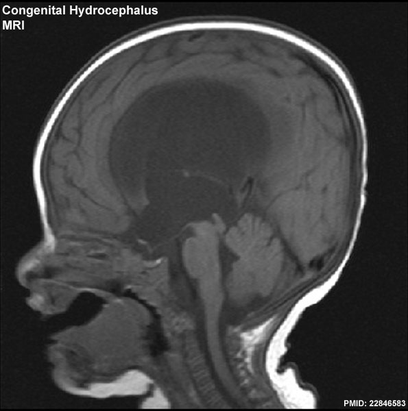 | 595 × 600 (43 KB) | Z8600021 (talk | contribs) | ==Congenital hydrocephalus (MRI)== Sagittal T1-weighted magnetic resonance image demonstrating obstructive hydrocephalus due to congenital aqueductal stenosis. ===Reference=== Bokhari and Baeesa Journal of Medical Case Reports 2012 6:222 doi:10.1186 |
You cannot overwrite this file.
File usage
The following 8 pages use this file:
- Abnormal Development - Congenital Hydrocephalus
- BGDA Lecture - Development of the Nervous System
- BGDA Practical 12 - Abnormalities
- BGD Lecture - Face and Ear Development
- BGD Tutorial - Applied Embryology and Teratology
- Lecture - 2016 Course Introduction
- Lecture - 2017 Course Introduction
- Template:Victoria abnormal data table 2004