File:Mouse 3D Heart external E8.5-14.5.jpeg: Difference between revisions
No edit summary |
No edit summary |
||
| Line 71: | Line 71: | ||
Three-dimensional reconstruction of gene expression patterns during cardiac development. Soufan AT, Ruijter JM, van den Hoff MJ, de Boer PA, Hagoort J, Moorman AF. Physiol Genomics. 2003 May 13;13(3):187-95. Review. [http://www.ncbi.nlm.nih.gov/pubmed/12746463 PMID: 12746463] | Three-dimensional reconstruction of gene expression patterns during cardiac development. Soufan AT, Ruijter JM, van den Hoff MJ, de Boer PA, Hagoort J, Moorman AF. Physiol Genomics. 2003 May 13;13(3):187-95. Review. [http://www.ncbi.nlm.nih.gov/pubmed/12746463 PMID: 12746463] | ||
[[Category:Heart]] | |||
Revision as of 22:53, 16 August 2009
Mouse 3D Heart external E8.5-14.5
Reconstructions of the myocardium of embryonic mouse hearts ranging from embryonic day 8.5 to 14.5 (ED 8.5 to 14.5).
All hearts are drawn at the same magnification (grid size: 500 x 500 µm). The increase of the myocardium volume is plotted as a curved line (y-axis in mm3). Note the similarity between the duplicate hearts at ED 9.5 and 10.5.
Fig. 2. H71230363002.jpeg
| Species | Stage | |||||||||||||||
| Human | Days | 20 | 22 | 24 | 28 | 30 | 33 | 36 | 40 | 42 | 44 | 48 | 52 | 54 | 55 | 58 |
| Mouse | Days | 9 | 9.5 | 10 | 10.5 | 11 | 11.5 | 12 | 12.5 | 13 | 13.5 | 14 | 14.5 | 15 | 15.5 | 16 |
Image (used with permission) from the paper Soufan AT, Ruijter JM, van den Hoff MJ, de Boer PA, Hagoort J, Moorman AF. Three-dimensional reconstruction of gene expression patterns during cardiac development. Physiol Genomics. 2003 May 13;13(3):187-95. (Physiol Genomics paper)
Three-dimensional reconstruction of gene expression patterns during cardiac development. Soufan AT, Ruijter JM, van den Hoff MJ, de Boer PA, Hagoort J, Moorman AF. Physiol Genomics. 2003 May 13;13(3):187-95. Review. PMID: 12746463
File history
Click on a date/time to view the file as it appeared at that time.
| Date/Time | Thumbnail | Dimensions | User | Comment | |
|---|---|---|---|---|---|
| current | 20:48, 16 August 2009 | 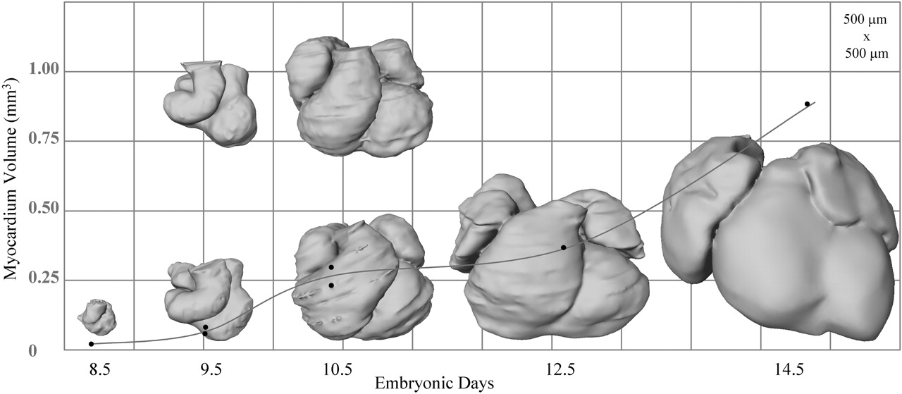 | 1,280 × 559 (90 KB) | S8600021 (talk | contribs) | Mouse 3D Heart external E8.5-14.5 Reconstructions of the myocardium of embryonic mouse hearts ranging from embryonic day 8.5 to 14.5 (ED 8.5 to 14.5). All hearts are drawn at the same magnification (grid size: 500 x 500 µm). The increase of the myocard |
You cannot overwrite this file.
File usage
The following 3 pages use this file: