File:Fetal integumentary histology 01.jpg: Difference between revisions
No edit summary |
m (→Reference) |
||
| Line 18: | Line 18: | ||
<pubmed>19701759</pubmed>| [http://www.ncbi.nlm.nih.gov/pmc/articles/PMC2799629 PMC2799629] | [http://www.springerlink.com/content/lv415257322x8247/fulltext.html Arch Dermatol Res] | <pubmed>19701759</pubmed>| [http://www.ncbi.nlm.nih.gov/pmc/articles/PMC2799629 PMC2799629] | [http://www.springerlink.com/content/lv415257322x8247/fulltext.html Arch Dermatol Res] | ||
====Copyright==== | |||
© Coolen NA, Schouten KC, Middelkoop E, Ulrich MM. 2009 Open Access - This article is distributed under the terms of the Creative Commons Attribution Noncommercial License which permits any noncommercial use, distribution, and reproduction in any medium, provided the original author(s) and source are credited. | © Coolen NA, Schouten KC, Middelkoop E, Ulrich MM. 2009 Open Access - This article is distributed under the terms of the Creative Commons Attribution Noncommercial License which permits any noncommercial use, distribution, and reproduction in any medium, provided the original author(s) and source are credited. | ||
| Line 24: | Line 24: | ||
Original file name: Fig. 1 403_2009_989_Fig1_HTML.gif (original figure increased in size) | Original file name: Fig. 1 403_2009_989_Fig1_HTML.gif (original figure increased in size) | ||
{{Footer}} | |||
[[Category:Integumentary]] [[Category:Histology]] [[Category:Human]] | [[Category:Integumentary]] [[Category:Histology]] [[Category:Human]] | ||
Revision as of 10:30, 10 October 2017
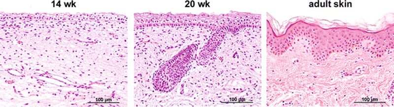
Fetal Integumentary Histology
- 14 weeks - the epidermis consisted of a basal layer, an intermediate cell layer and periderm.
- 20 weeks - the number of intermediate cells layers was increased and developing hair follicles were visible.
- adult skin - contained basal, spinous, granular and cornified layers.
Note these ages appear to be LMP - This study gestational ages were estimated by menstrual age and ranged from 13 to 22 weeks. The embryo gestational ages and numbers were as follows: gestational age 13 weeks (1), 14 weeks (1), 16 weeks (1), 17 weeks (2), 18 weeks (2), 19 weeks (4), 20 weeks (4), 21 weeks (3), 22 weeks (3).
Scale bars 100 μm
In early gestation (13–14 weeks), fetal epidermis contained a basal layer, one or two intermediate layers and a periderm. At 14 weeks, the fetal dermis consisted of a finely fibrillar dermis that contained many cells. From 16 weeks of gestation, hair pegs were visible that projected into the dermis. The dermis was organized into two regions: an upper fibrillar papillary region and a deep reticular region that contained larger fibers. During further development, the number of epidermal cell layers increased and the hair pegs matured into hair follicles. Developing eccrine sweat glands were detected from week 21.
Reference
<pubmed>19701759</pubmed>| PMC2799629 | Arch Dermatol Res
Copyright
© Coolen NA, Schouten KC, Middelkoop E, Ulrich MM. 2009 Open Access - This article is distributed under the terms of the Creative Commons Attribution Noncommercial License which permits any noncommercial use, distribution, and reproduction in any medium, provided the original author(s) and source are credited.
Original file name: Fig. 1 403_2009_989_Fig1_HTML.gif (original figure increased in size)
Cite this page: Hill, M.A. (2024, June 3) Embryology Fetal integumentary histology 01.jpg. Retrieved from https://embryology.med.unsw.edu.au/embryology/index.php/File:Fetal_integumentary_histology_01.jpg
- © Dr Mark Hill 2024, UNSW Embryology ISBN: 978 0 7334 2609 4 - UNSW CRICOS Provider Code No. 00098G
File history
Click on a date/time to view the file as it appeared at that time.
| Date/Time | Thumbnail | Dimensions | User | Comment | |
|---|---|---|---|---|---|
| current | 00:44, 10 October 2010 | 800 × 219 (74 KB) | S8600021 (talk | contribs) | ==Histology of 14 and 20 weeks’ gestation fetal skin and of adult skin== ==Fetal integumentary histology== * At 14 weeks, the epidermis consisted of a basal layer, an intermediate cell layer and periderm. * At 20 weeks, the number of intermediate cell |
You cannot overwrite this file.