File:Human fetal temporal bone and mandible 01.jpg: Difference between revisions
From Embryology
mNo edit summary |
mNo edit summary |
||
| (3 intermediate revisions by the same user not shown) | |||
| Line 4: | Line 4: | ||
===Temporal Bone=== | |||
'''Temporal styloid''' process growth can also be noted. | '''Temporal styloid''' process growth can also be noted. | ||
* | * pointed piece of bone extending from the temporal bone just below the ear. | ||
* anchor point for several muscles associated with the tongue and larynx. | |||
===Mandible=== | |||
* develops beside the Meckel's cartilage template. | |||
* bone formation by intramembranous ossification. | |||
* at birth not completely ossified. | |||
* there is a sex difference in overall growth. | |||
:'''Links:''' [[Musculoskeletal_System_-_Skull_Development|Skull Development]] | |||
===Reference=== | |||
{{Virginia Diewert images}} | {{Virginia Diewert images}} | ||
{{ | {{Footer}} | ||
[[Category:Human Fetus]] [[Category:Musculoskeletal]] [[Category:Bone]] [[Category:Head]] [[Category:Skull]] | [[Category:Human Fetus]] [[Category:Musculoskeletal]] [[Category:Bone]] [[Category:Head]] [[Category:Skull]] | ||
Latest revision as of 17:14, 24 July 2017
Human Temporal Bone and Mandible
Image shows growth of both bones from the end of the embryonic period (week 8, GA week 10) through the fetal period of development (to 9 months).
Temporal Bone
Temporal styloid process growth can also be noted.
- pointed piece of bone extending from the temporal bone just below the ear.
- anchor point for several muscles associated with the tongue and larynx.
Mandible
- develops beside the Meckel's cartilage template.
- bone formation by intramembranous ossification.
- at birth not completely ossified.
- there is a sex difference in overall growth.
- Links: Skull Development
Reference
Image Source: Prof Virginia Diewert
Cite this page: Hill, M.A. (2024, June 1) Embryology Human fetal temporal bone and mandible 01.jpg. Retrieved from https://embryology.med.unsw.edu.au/embryology/index.php/File:Human_fetal_temporal_bone_and_mandible_01.jpg
- © Dr Mark Hill 2024, UNSW Embryology ISBN: 978 0 7334 2609 4 - UNSW CRICOS Provider Code No. 00098G
File history
Click on a date/time to view the file as it appeared at that time.
| Date/Time | Thumbnail | Dimensions | User | Comment | |
|---|---|---|---|---|---|
| current | 14:42, 15 May 2013 | 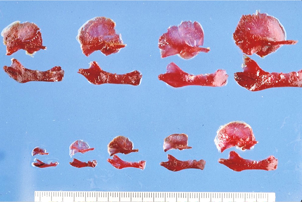 | 1,200 × 805 (170 KB) | Z8600021 (talk | contribs) | ==Human Temporal Bone and Mandible== Image shows growth of both bones from the end of the embryonic period (week 8) through the fetal period of development (to 9 months). ---- Image Source: Prof Virginia Diewert {{Template:Footer}} [[Category:Human... |
You cannot overwrite this file.
File usage
The following page uses this file: