File:Crowder1957 plate02.jpg: Difference between revisions
m (→Plate 2) |
m (→Plate 2) |
||
| Line 1: | Line 1: | ||
==Plate 2== | ==Plate 2== | ||
Fig. 12. Area of rectangle in figure ll, .~;lmwin_:z cells of | Fig. 12. Area of rectangle in figure ll, .~;lmwin_:z cells of llnwni;ui'.~'. capsule. At arrow .-1. cells appear to he migrating into |]1I3!iL‘|1Cll}’!11C dorsal to adrenal primordium. .-\t arrmv b’. they appear to he migrating intn the adrenal and gmiadal prinmr. dia. Small cells (C) with elongated nuclei are seen in the ten- t1'nl;1teI':Il pnrlinn of the gland. .\'n. pl. I4—3—5'. >1 ‘gun. in, ad- renal grucwe. | ||
llnwni;ui'.~'. capsule. At arrow .-1. cells appear to he migrating | |||
into |]1I3!iL‘|1Cll}’!11C dorsal to adrenal primordium. .-\t arrmv b’. | |||
they appear to he migrating intn the adrenal and gmiadal prinmr. | |||
dia. Small cells (C) with elongated nuclei are seen in the ten- | |||
t1'nl;1teI':Il pnrlinn of the gland. .\'n. pl. I4—3—5'. >1 ‘gun. in, ad- | |||
renal grucwe. | |||
Fig. 13. Frum the same emhryn as figure ll. 2:) microns | Fig. 13. Frum the same emhryn as figure ll. 2:) microns c:iudal to section slimwi in figure 1.3. >-(($00. 1, type C—| cells: 2. type (LII Cell; (I. 5. c‘., primordial sex cell. | ||
c:iudal to section slimwi in figure 1.3. >-(($00. 1, type C—| cells: 2. | |||
type (LII Cell; (I. 5. c‘., primordial sex cell. | |||
Fig. 14. From :m emhr_\~'n of 3 weeks. Primordia of adrenal and synipatlietie ganglion (I3) are relatively larger. compared to | Fig. 14. From :m emhr_\~'n of 3 weeks. Primordia of adrenal and synipatlietie ganglion (I3) are relatively larger. compared to inesuneplirus, than in figure ll (pl. I). Adrenal primurdium in direct euntinuity with eelnmie epithelium. Nu. 792, 10-1-3. I-luriznn xvi. >< mu. 5' mm. .1. aurta: 7. adrenal primurditim: 1.1 (arruw), hand of cells nf mesnnephrie origin (‘type C-ll). | ||
inesuneplirus, than in figure ll (pl. I). Adrenal primurdium in | |||
direct euntinuity with eelnmie epithelium. Nu. 792, 10-1-3. | |||
I-luriznn xvi. >< mu. 5' mm. .1. aurta: 7. adrenal primurditim: 1.1 | |||
(arruw), hand of cells nf mesnnephrie origin (‘type C-ll). | |||
Fig. 15. From a section near that slirm'n in figure 14. Type | Fig. 15. From a section near that slirm'n in figure 14. Type C—ll cells (2) appear to he migrating along ventral .~‘.urf:iee of primurdium from Hnxviiizilfs capsule. lnvading neural elements (armw) from s}'mp:Itlietie chain spreading widely amnng the cells of the primnrdium have disttirhed its lmniugeneity. D, s_x-'i1ip;tt|ietic nerve. >-: _=;0n. | ||
C—ll cells (2) appear to he migrating along ventral .~‘.urf:iee of | |||
primurdium from Hnxviiizilfs capsule. lnvading neural elements (armw) from s}'mp:Itlietie chain spreading widely amnng | |||
the cells of the primnrdium have disttirhed its lmniugeneity. D, | |||
s_x-'i1ip;tt|ietic nerve. >-: _=;0n. | |||
{{Crowder1957 figures}} | {{Crowder1957 figures}} | ||
[[Category:Carnegie Stage 15]][[Category:Week 5]][[Category:Carnegie Embryo | [[Category:Carnegie Stage 15]][[Category:Week 5]][[Category:Carnegie Embryo 792]][[Category:Carnegie Embryo 721]] | ||
Latest revision as of 23:03, 21 June 2017
Plate 2
Fig. 12. Area of rectangle in figure ll, .~;lmwin_:z cells of llnwni;ui'.~'. capsule. At arrow .-1. cells appear to he migrating into |]1I3!iL‘|1Cll}’!11C dorsal to adrenal primordium. .-\t arrmv b’. they appear to he migrating intn the adrenal and gmiadal prinmr. dia. Small cells (C) with elongated nuclei are seen in the ten- t1'nl;1teI':Il pnrlinn of the gland. .\'n. pl. I4—3—5'. >1 ‘gun. in, ad- renal grucwe.
Fig. 13. Frum the same emhryn as figure ll. 2:) microns c:iudal to section slimwi in figure 1.3. >-(($00. 1, type C—| cells: 2. type (LII Cell; (I. 5. c‘., primordial sex cell.
Fig. 14. From :m emhr_\~'n of 3 weeks. Primordia of adrenal and synipatlietie ganglion (I3) are relatively larger. compared to inesuneplirus, than in figure ll (pl. I). Adrenal primurdium in direct euntinuity with eelnmie epithelium. Nu. 792, 10-1-3. I-luriznn xvi. >< mu. 5' mm. .1. aurta: 7. adrenal primurditim: 1.1 (arruw), hand of cells nf mesnnephrie origin (‘type C-ll).
Fig. 15. From a section near that slirm'n in figure 14. Type C—ll cells (2) appear to he migrating along ventral .~‘.urf:iee of primurdium from Hnxviiizilfs capsule. lnvading neural elements (armw) from s}'mp:Itlietie chain spreading widely amnng the cells of the primnrdium have disttirhed its lmniugeneity. D, s_x-'i1ip;tt|ietic nerve. >-: _=;0n.
- Links: fig 1 | fig 2 | fig 3 | fig 4 | fig 5 | fig 6 | fig 7 | fig 1-7 | plate 1 | plate 2 | plate 3 | 1957 Crowder | Adrenal Development
References
Crowder RE. The development of the adrenal gland in man, with special reference to origin and ultimate location of cell types and evidence in favor of the "cell migration" theory. (1957) Contrib. Embryol., Carnegie Inst. Wash. 36, 193-210.
Cite this page: Hill, M.A. (2024, June 2) Embryology Crowder1957 plate02.jpg. Retrieved from https://embryology.med.unsw.edu.au/embryology/index.php/File:Crowder1957_plate02.jpg
- © Dr Mark Hill 2024, UNSW Embryology ISBN: 978 0 7334 2609 4 - UNSW CRICOS Provider Code No. 00098G
File history
Click on a date/time to view the file as it appeared at that time.
| Date/Time | Thumbnail | Dimensions | User | Comment | |
|---|---|---|---|---|---|
| current | 21:16, 21 June 2017 | 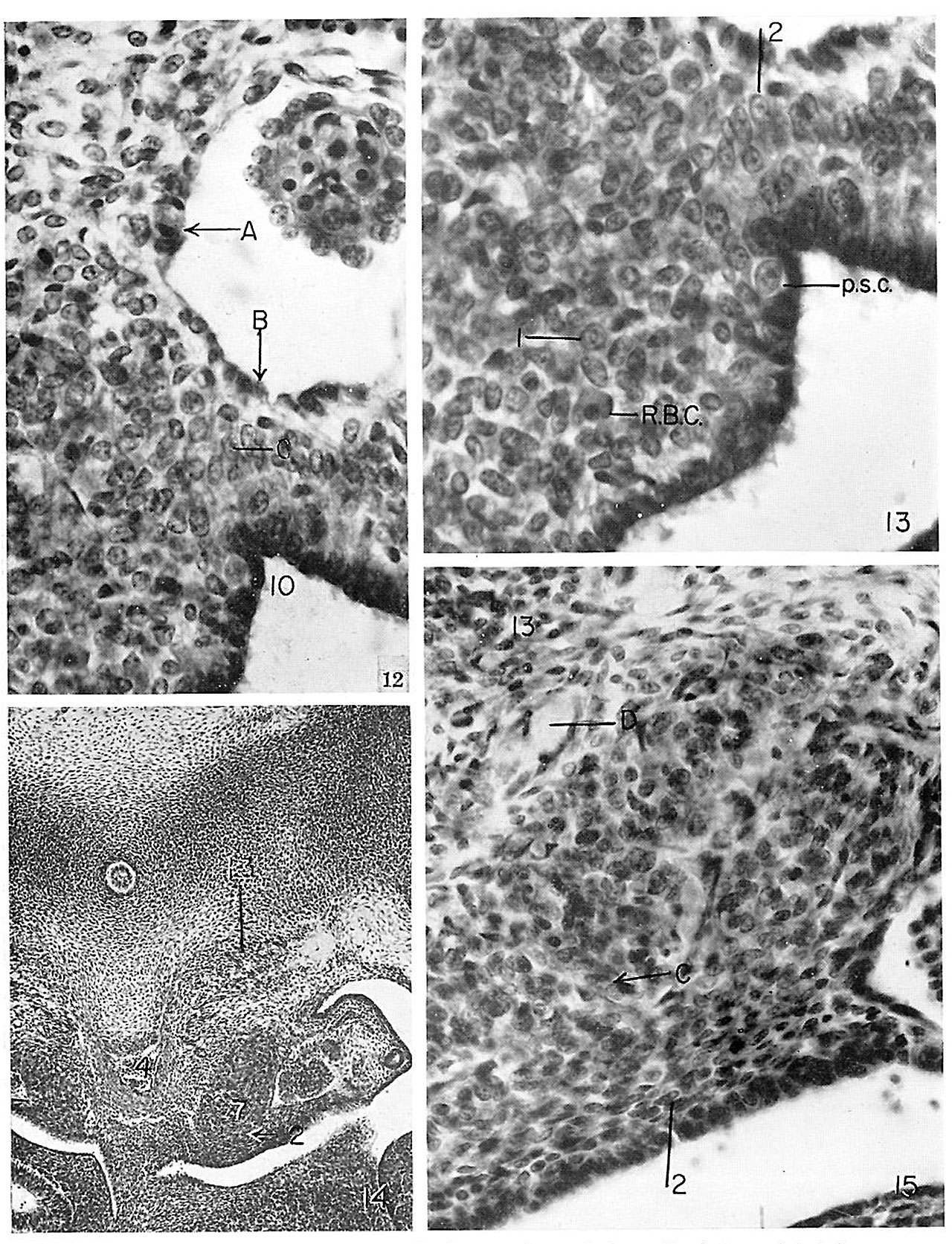 | 1,280 × 1,673 (645 KB) | Z8600021 (talk | contribs) | |
| 21:14, 21 June 2017 |  | 2,195 × 3,167 (1.38 MB) | Z8600021 (talk | contribs) | ==Plate 2== {{Crowder1957 figures}} |
You cannot overwrite this file.
File usage
The following page uses this file: