Category:Neural Crest: Difference between revisions
From Embryology
mNo edit summary |
mNo edit summary |
||
| Line 4: | Line 4: | ||
{{Neural Crest Links}} | {{Neural Crest Links}} | ||
[[Category:Ectoderm]] | [[Category:Ectoderm]] | ||
Revision as of 15:44, 20 May 2016
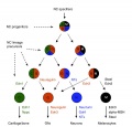
This Embryology category shows pages and media related to neural crest development. This special population of cells is ectoderm in origin and forms many different types of tissue throughout the embryo.
| Neural Crest Links: neural crest | Lecture - Early Neural | Lecture - Neural Crest Development | Lecture Movie | Schwann cell | adrenal | melanocyte | peripheral nervous system | enteric nervous system | cornea | cranial nerve neural crest | head | skull | cardiac neural crest | Nicole Le Douarin | Neural Crest Movies | neural crest abnormalities | Category:Neural Crest | |||
|
Subcategories
This category has the following 7 subcategories, out of 7 total.
Pages in category 'Neural Crest'
The following 109 pages are in this category, out of 109 total.
2
C
- Template:Cardiac neural crest
- Chicken Development
- Chicken Neural Crest Migration Movie 1
- Chicken Neural Crest Migration Movie 2
- Chicken Neural Crest Migration Movie 3
- Chicken Neural Crest Migration Movie 4
- Chicken Neural Crest Migration Movie 5
- Chicken Neural Crest Migration Movie 6
- Chicken Neural Crest Migration Movie 7
- Template:Chicken neural crest movies
- Template:Cornea
- Template:Cranial nerve neural crest
- Template:Cranial Neural Crest Timeline table
D
E
H
M
- Template:Mandible
- Template:Meckel’s cartilage
- Template:Megacolon
- Template:Melanocyte
- Template:Merkel cell
- Mouse Cranial Neural Crest Migration Movie
- Mouse Melanoblast Migration Movie
- Template:Mouse neural crest pax7 links
- Movie - Chicken Neural Crest Migration 01
- Movies
- Movies - Chicken Neural Crest
- Template:Myenteric plexus
N
- Template:Neural crest
- Neural Crest - Cardiac
- Neural Crest - Cranial Nerve Development
- Neural Crest - Cranial Nerves
- Neural Crest - Enteric Nervous System
- Neural Crest - Melanocyte Development
- Neural Crest - Peripheral Nervous System
- Neural Crest - Schwann Cell Development
- Template:Neural crest abnormalities
- Template:Neural Crest collapsible table
- Neural Crest Development
- Template:Neural Crest Links
- Template:Neural crest origin collapsetable1
- Template:Neural crest origin table1
- Neural Crest System - Abnormalities
- Template:Neural Crest table
- Template:Neural Crest Vignette
P
- Paper - 1879 The Morphology of the Vertebrate Olfactory Organ
- Paper - A contribution to the histogenesis of the sympathetic nervous system (1909)
- Paper - A nerve growth-stimulating factor isolated from sarcomas 37 and 180
- Paper - Observations on the neural crest of a ten-somite human embryo (1939)
- Paper - Problems concerning the origin and development of the neural crest and cranial ganglia in the vertebrates (1928)
- Paper - The development of the adrenal gland in man (1957)
- Paper - The Development of the Cranial and Spinal Nerves in the Occipital Region of the Human Embryo
- Paper - The development of the sympathetic nervous system in mammals
- Paper - The development of the sympathetic nervous system in mammals (1910)
- Paper - The development of the sympathetic nervous system in man
- Paper - The development of the sympathetic system in birds (1910)
- Paper - The origin of the neural crest
- Paper - The Peripheral Nervous System in the Human Embryo at the End of the First Month (10 mm)
- Paper - The role of the vagi in the development of the sympathetic nervous system (1909)
- Template:Peripheral nervous system
- Template:Peripheral Nervous System
- Template:Pharyngeal arch
- Template:PNS
R
Media in category 'Neural Crest'
The following 93 files are in this category, out of 93 total.
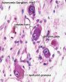 Autonomic ganglion histology 01.jpg 641 × 800; 56 KB
 Bailey366.jpg 558 × 633; 97 KB
 Bailey391.jpg 633 × 485; 56 KB
 BaxterBoyd1939-fig01.jpg 361 × 736; 43 KB
 BaxterBoyd1939-fig02.jpg 459 × 851; 65 KB
 BaxterBoyd1939-fig03.jpg 732 × 1,000; 170 KB
 BaxterBoyd1939-fig04.jpg 642 × 615; 109 KB
 BaxterBoyd1939-fig05.jpg 1,000 × 864; 263 KB
 BaxterBoyd1939-fig06.jpg 489 × 917; 141 KB
 BaxterBoyd1939-fig07.jpg 795 × 917; 242 KB
 BaxterBoyd1939-plate01.jpg 1,680 × 2,400; 786 KB
 BaxterBoyd1939-plate02.jpg 1,681 × 2,400; 860 KB
 BaxterBoyd1939-text-fig01.jpg 1,283 × 1,000; 138 KB
 BaxterBoyd1939-text-fig02.jpg 1,200 × 861; 133 KB
 Cardiac Neural Crest Migration.jpg 1,517 × 1,116; 122 KB
 Carnegie stage 13 caudal trunk.jpg 400 × 625; 53 KB
 Chicken-neural-crest-migration-01.jpg 600 × 432; 37 KB
 Chicken-neural-crest-migration-02.jpg 600 × 419; 43 KB
 Chicken-neural-crest-migration-03.jpg 600 × 346; 53 KB
 Chicken-neural-crest-migration-04.jpg 600 × 397; 67 KB
 Chicken-neural-crest-migration-05.jpg 600 × 586; 66 KB
 Chicken-neural-crest-migration-06.jpg 600 × 398; 40 KB
 Chicken-neural-crest-migration-07.jpg 600 × 453; 45 KB
 Childhood cancer survival rates.jpg 536 × 335; 25 KB
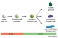 Cochlea glial lineage cartoon.jpg 1,000 × 651; 52 KB
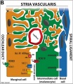 Cochlea stria vascularis cartoon 03.jpg 694 × 800; 125 KB
 Cranial neural crest skeletal fate 01.jpg 800 × 633; 59 KB
 Eye-neural crest signaling.jpg 946 × 886; 436 KB
 Gray0847.jpg 559 × 900; 155 KB
 Gray0848.jpg 800 × 935; 289 KB
 Gray0849.jpg 800 × 885; 258 KB
 Hindbrain neural crest migration.jpg 450 × 545; 48 KB
 Human Carnegie stage 13 GJA1 expression.jpg 706 × 470; 96 KB
 Human Carnegie stage 13 SOX11 MAZ GJA1 expression.jpg 777 × 1,000; 192 KB
 Human Carnegie stage 13 stem cell markers.jpg 657 × 585; 78 KB
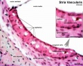
 Human cochlea stria vascularis 01.jpg 1,854 × 1,806; 754 KB
 Human neural crest cell migration-in vitro.jpg 1,280 × 959; 163 KB
 Human-adrenal gland 01.jpg 596 × 392; 65 KB
 Keith1902 fig035.jpg 800 × 633; 125 KB
 LeDouarin.jpg 156 × 198; 19 KB
 Lens-neural crest signaling 01.jpg 300 × 400; 24 KB
 Lens-neural crest signaling 02.jpg 521 × 522; 22 KB
 Megacolon stoma.gif 200 × 202; 6 KB
 Melanoblast migration.png 600 × 210; 40 KB
 Melanocyte development cartoon.jpg 1,280 × 1,086; 176 KB
 Merkel cell EM 01.jpg 984 × 738; 209 KB
 Merkel cell EM 02.jpg 984 × 685; 166 KB
 Model neural crest mesenchymal condensation.jpg 818 × 1,200; 131 KB
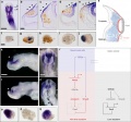
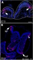
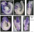 Mouse E9.5 neural crest - Crabp1, Sox10, Pax3.jpg 1,323 × 1,233; 193 KB
 Mouse E9.5 neural crest - Crabp1.jpg 2,489 × 626; 217 KB
 Mouse E9.5 neural crest beta actin KO.jpg 2,722 × 2,573; 732 KB
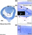 Mouse eye E18.jpg 884 × 977; 169 KB
 Mouse eye neural crest cornea 01.jpg 500 × 256; 34 KB
 Mouse eye neural crest cornea 02.jpg 800 × 655; 121 KB
 Mouse eye neural crest.jpg 1,086 × 1,509; 483 KB
 Mouse eye TGF-beta model.jpg 1,000 × 519; 88 KB
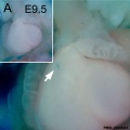
 Mouse head E9-neural crest GFP.jpg 539 × 543; 56 KB
 Mouse head-neural crest 01.jpg 900 × 339; 48 KB
 Mouse melanoblast distribution 01.jpg 697 × 1,000; 192 KB
 Mouse melanoblast distribution 02.jpg 777 × 1,121; 129 KB
 Mouse melanoblast distribution 03.jpg 751 × 1,051; 150 KB
 Mouse melanoblast distribution 04.jpg 761 × 1,128; 177 KB
 Mouse melanoblast distribution 05.jpg 761 × 540; 94 KB
 Mouse melanoblast distribution 06.jpg 761 × 524; 74 KB
 Mouse organ of corti 04.jpg 1,280 × 1,024; 202 KB
 Mouse pax7 heart 01.jpg 561 × 560; 34 KB
 Mouse pax7 limb 01.jpg 1,320 × 549; 112 KB
 Mouse pax7 neural fold 01.jpg 513 × 915; 100 KB
 Mouse pax7 trunk neural crest 01.jpg 881 × 691; 158 KB
 Mouse pax7 trunk neural crest 02.jpg 881 × 691; 171 KB
 Mouse-E10.5 ganglia Sox10.jpg 540 × 685; 51 KB
 Mouse-E10.5-Sox10.jpg 400 × 463; 24 KB
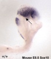 Mouse-E8.5-Sox10.jpg 400 × 463; 20 KB
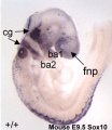 Mouse-E9.5-Sox10.jpg 400 × 463; 30 KB
 Mouse-melanoblast migration icon.jpg 450 × 450; 59 KB
 Mouse-neural crest Sox10 E10.5.jpg 1,000 × 420; 55 KB
 Neural - cranial nerves.jpg 800 × 447; 72 KB
 Neural crest cell enteric sorting.jpg 800 × 849; 61 KB
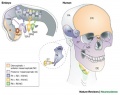
 Neural crest contribution.jpg 1,280 × 701; 64 KB
 Neural crest formation stages 01.jpg 1,200 × 728; 124 KB
 Neural crest precursor differentiation.jpg 600 × 576; 42 KB
 Neuroblastoma.jpg 640 × 426; 31 KB
 Smooth muscle histology 002.jpg 600 × 750; 112 KB
 Stage 22 image 152.jpg 1,000 × 670; 127 KB
 Stage 22 image 153.jpg 1,000 × 662; 191 KB
 Stage 22 image 155.jpg 1,000 × 672; 239 KB
 Stage11 sem21.jpg 600 × 447; 79 KB
 Trunk neural crest migration.jpg 1,280 × 963; 220 KB
 Week10 adrenal.jpg 600 × 564; 48 KB
 Wen1928-Fig10.jpg 495 × 1,200; 108 KB
 Zebrafish melanocyte development model.jpg 484 × 277; 40 KB
 Zebrafish neural crest model.jpg 600 × 480; 61 KB
 Zebrafish skull neural crest.jpg 815 × 933; 187 KB


