File:Pincus1935-plate01.jpg: Difference between revisions
m (→Plate 1) |
mNo edit summary |
||
| (3 intermediate revisions by the same user not shown) | |||
| Line 1: | Line 1: | ||
==Plate 1== | ==Plate 1== | ||
<gallery> | |||
File:Pincus1935-fig01.jpg|FIG. 1. Ovarian egg obtained by puncture of a follicle from the ovary of an unmated rabbit. | |||
File:Pincus1935-fig02.jpg|FIG. 2. Ovarian egg obtained by puncture of a follicle from the ovary of a doe mated 2 hours previously. The chromatin material condenses to tetrads. The vesicular membrane is still present. | |||
File:Pincus1935-fig05.jpg|FIG. 5. Ovarian egg from a doe mated 6 hours previously. The first polar spindle begins to form. | |||
File:Pincus1935-fig06.jpg|FIG. 6. Ovarian egg from a doe mated 8 hours previously. The first polar body has been given off. | |||
File:Pincus1935-fig09.jpg|FIG. 9. Ovarian egg from a doe which had received 2 cc. thyroxin intravenously. Tetrads have formed in a vesicular nucleus. | |||
File:Pincus1935-fig10.jpg|FIG. 10. Ovarian egg from unmated doe, inseminated with normal sperm in vitro. Sperm penetration has occurred. Note sperm head at lower right periphery. | |||
</gallery> | |||
{{Pincus1935 figures}} | |||
{{ | |||
Latest revision as of 19:38, 22 April 2016
Plate 1
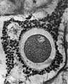
FIG. 1. Ovarian egg obtained by puncture of a follicle from the ovary of an unmated rabbit.
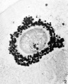
FIG. 2. Ovarian egg obtained by puncture of a follicle from the ovary of a doe mated 2 hours previously. The chromatin material condenses to tetrads. The vesicular membrane is still present.
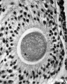
FIG. 5. Ovarian egg from a doe mated 6 hours previously. The first polar spindle begins to form.
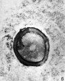
FIG. 6. Ovarian egg from a doe mated 8 hours previously. The first polar body has been given off.
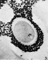
FIG. 9. Ovarian egg from a doe which had received 2 cc. thyroxin intravenously. Tetrads have formed in a vesicular nucleus.
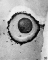
FIG. 10. Ovarian egg from unmated doe, inseminated with normal sperm in vitro. Sperm penetration has occurred. Note sperm head at lower right periphery.


| Historic Disclaimer - information about historic embryology pages |
|---|
 |
Reference
Pincus G. and Enzmann EV. The comparative behavior of mammalian eggs in vivo and in vitro. (1935) J Exp Med. 62(5): 665-75. PMID 19870440
Cite this page: Hill, M.A. (2024, June 2) Embryology Pincus1935-plate01.jpg. Retrieved from https://embryology.med.unsw.edu.au/embryology/index.php/File:Pincus1935-plate01.jpg
- © Dr Mark Hill 2024, UNSW Embryology ISBN: 978 0 7334 2609 4 - UNSW CRICOS Provider Code No. 00098G
File history
Click on a date/time to view the file as it appeared at that time.
| Date/Time | Thumbnail | Dimensions | User | Comment | |
|---|---|---|---|---|---|
| current | 09:25, 15 November 2015 | 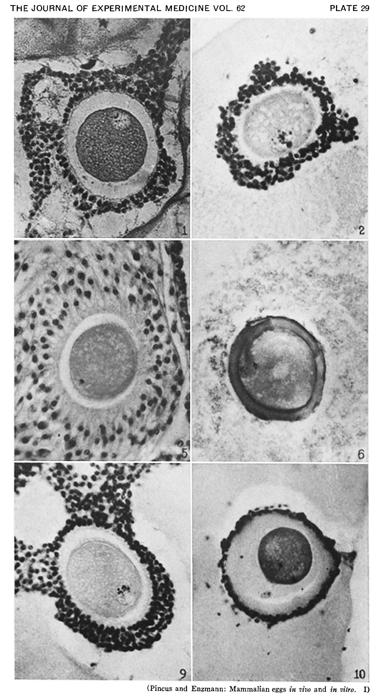 | 1,300 × 2,326 (463 KB) | Z8600021 (talk | contribs) |
You cannot overwrite this file.
File usage
The following page uses this file: