File:West09.jpg: Difference between revisions
mNo edit summary |
mNo edit summary |
||
| Line 1: | Line 1: | ||
==Fig. 9. Otocyst and Neural Tube== | ==Fig. 9. Otocyst and Neural Tube== | ||
The neuromeres resemble closely those described by other observers, but they show more clearly than in other specimens (Text-fig. 9); there are ten of these segments which involve the lateral wall of the brain. The 2nd, 4th, 6th and 7th involve also the flor, to which they give an irregular outline;itisthe grooves between the projections of the floor caused by these neuromeres, and especially the 4th, which are referred to by Thompson (1907), Bremer (1906), and van den Broek (1911),as having given rise to a difficulty in their interpretation, and van den Broek suggested that they might have some association with the Vth and VIlIth ganglia, which is indirectly true in view of the relation of these ganglia to the neuromeres which are responsible for the production of these grooves. The ganglion of the trigeminal nerve lies lateral to segments 2 and 3; the acoustico-facial ganglion is lateral to segment 4; the otocyst is lateral to segment 5; the glossopharyngeal ganglion is lateral to segment 7, and the vagus ganglion is lateral to segments 7 and 8 (Text-figs. 8 and 9), but the glossopharyngeal and vagus ganglia are difficult to define and appear only as rather diffuse clumps of cels.The first somite is opposite the 8th neuromere, the 2nd somite is opposite the 9th neuromere, and the 3rd is opposite the 9th and 1oth neuromeres and the intervening groove. | |||
Revision as of 12:16, 17 February 2016
Fig. 9. Otocyst and Neural Tube
The neuromeres resemble closely those described by other observers, but they show more clearly than in other specimens (Text-fig. 9); there are ten of these segments which involve the lateral wall of the brain. The 2nd, 4th, 6th and 7th involve also the flor, to which they give an irregular outline;itisthe grooves between the projections of the floor caused by these neuromeres, and especially the 4th, which are referred to by Thompson (1907), Bremer (1906), and van den Broek (1911),as having given rise to a difficulty in their interpretation, and van den Broek suggested that they might have some association with the Vth and VIlIth ganglia, which is indirectly true in view of the relation of these ganglia to the neuromeres which are responsible for the production of these grooves. The ganglion of the trigeminal nerve lies lateral to segments 2 and 3; the acoustico-facial ganglion is lateral to segment 4; the otocyst is lateral to segment 5; the glossopharyngeal ganglion is lateral to segment 7, and the vagus ganglion is lateral to segments 7 and 8 (Text-figs. 8 and 9), but the glossopharyngeal and vagus ganglia are difficult to define and appear only as rather diffuse clumps of cels.The first somite is opposite the 8th neuromere, the 2nd somite is opposite the 9th neuromere, and the 3rd is opposite the 9th and 1oth neuromeres and the intervening groove.
| Historic Disclaimer - information about historic embryology pages |
|---|
 |
- Links: Fig 1 | Fig 2 | Fig 3 | Fig 4 | Fig 5 | Fig 6 | Fig 7 | Fig 8 | Fig 9 | Fig 10 | Fig 11 | Plate 1 | Plate 1 Fig 1 | Plate 1 Fig 2 | Plate 1 Fig 3 | Plate 1 Fig 4 | Plate 1 Fig 5 | Plate 1 Fig 6
Reference
West CM. A human embryo of twenty-five somites. (1937) J. Anat., 71(2): 169-200.1. PMID 17104635
Cite this page: Hill, M.A. (2024, June 1) Embryology West09.jpg. Retrieved from https://embryology.med.unsw.edu.au/embryology/index.php/File:West09.jpg
- © Dr Mark Hill 2024, UNSW Embryology ISBN: 978 0 7334 2609 4 - UNSW CRICOS Provider Code No. 00098G
Reference
West, CM. A Human Embryo of Twenty-five Somites. J. Anat.: 1937, 71(Pt 2);169-200.1 PMID 17104635
Cite this page: Hill, M.A. (2024, June 1) Embryology West09.jpg. Retrieved from https://embryology.med.unsw.edu.au/embryology/index.php/File:West09.jpg
- © Dr Mark Hill 2024, UNSW Embryology ISBN: 978 0 7334 2609 4 - UNSW CRICOS Provider Code No. 00098G
File history
Click on a date/time to view the file as it appeared at that time.
| Date/Time | Thumbnail | Dimensions | User | Comment | |
|---|---|---|---|---|---|
| current | 15:22, 28 January 2012 | 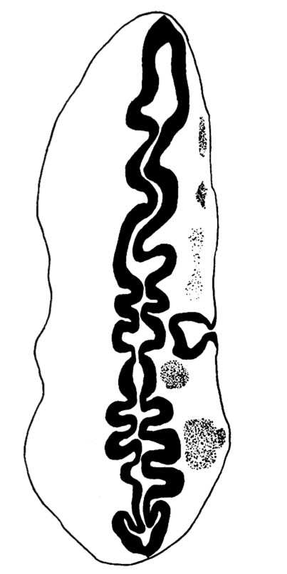 | 403 × 806 (44 KB) | S8600021 (talk | contribs) | ==Fig. 9 == {{Template:West1937}} {{Historic Disclaimer}} {{Historic Papers}} ===Reference=== <pubmed>17104635</pubmed>| [http://www.ncbi.nlm.nih.gov/pmc/articles/PMC1252340 PMC1252340] Category:Carnegie Stage 12 |
You cannot overwrite this file.
File usage
The following page uses this file: