File:Streeter1908-fig01.jpg: Difference between revisions
(Z8600021 uploaded a new version of File:Streeter1908-fig01.jpg) |
mNo edit summary |
||
| Line 1: | Line 1: | ||
==Fig. 1. Composite sagittal view of a 10 mm Human Embryo== | ==Fig. 1. Composite sagittal view of a 10 mm Human Embryo== | ||
(Huber collection No. 3), showing the rhombencephalon and the | (Huber collection No. 3), showing the rhombencephalon and the so-called rhombic grooves, which exist temporarily as transverse furrows in the floor of the ventricle. They are connected by the fifth, seventh, ninth and tenth nerves with the branchiai arches. The fourth groove (d.) is an exception and has no corresponding visceral nerve. Arising from it can be seen the sixth nerve, extending forward to reach the anlage of the external rectus muscle. | ||
{{Streeter1908 figures}} | {{Streeter1908 figures}} | ||
Latest revision as of 09:36, 15 September 2015
Fig. 1. Composite sagittal view of a 10 mm Human Embryo
(Huber collection No. 3), showing the rhombencephalon and the so-called rhombic grooves, which exist temporarily as transverse furrows in the floor of the ventricle. They are connected by the fifth, seventh, ninth and tenth nerves with the branchiai arches. The fourth groove (d.) is an exception and has no corresponding visceral nerve. Arising from it can be seen the sixth nerve, extending forward to reach the anlage of the external rectus muscle.
| Historic Disclaimer - information about historic embryology pages |
|---|
 |
| Online Editor | ||||||||||||||||||
|---|---|---|---|---|---|---|---|---|---|---|---|---|---|---|---|---|---|---|
|
Reference
Streeter GL. The peripheral nervous system in the human embryo at the end of the first month (10 mm) (1908) Amer. J Anat. 8(1): 285–302.
Cite this page: Hill, M.A. (2024, June 14) Embryology Streeter1908-fig01.jpg. Retrieved from https://embryology.med.unsw.edu.au/embryology/index.php/File:Streeter1908-fig01.jpg
- © Dr Mark Hill 2024, UNSW Embryology ISBN: 978 0 7334 2609 4 - UNSW CRICOS Provider Code No. 00098G
File history
Click on a date/time to view the file as it appeared at that time.
| Date/Time | Thumbnail | Dimensions | User | Comment | |
|---|---|---|---|---|---|
| current | 12:11, 14 September 2015 | 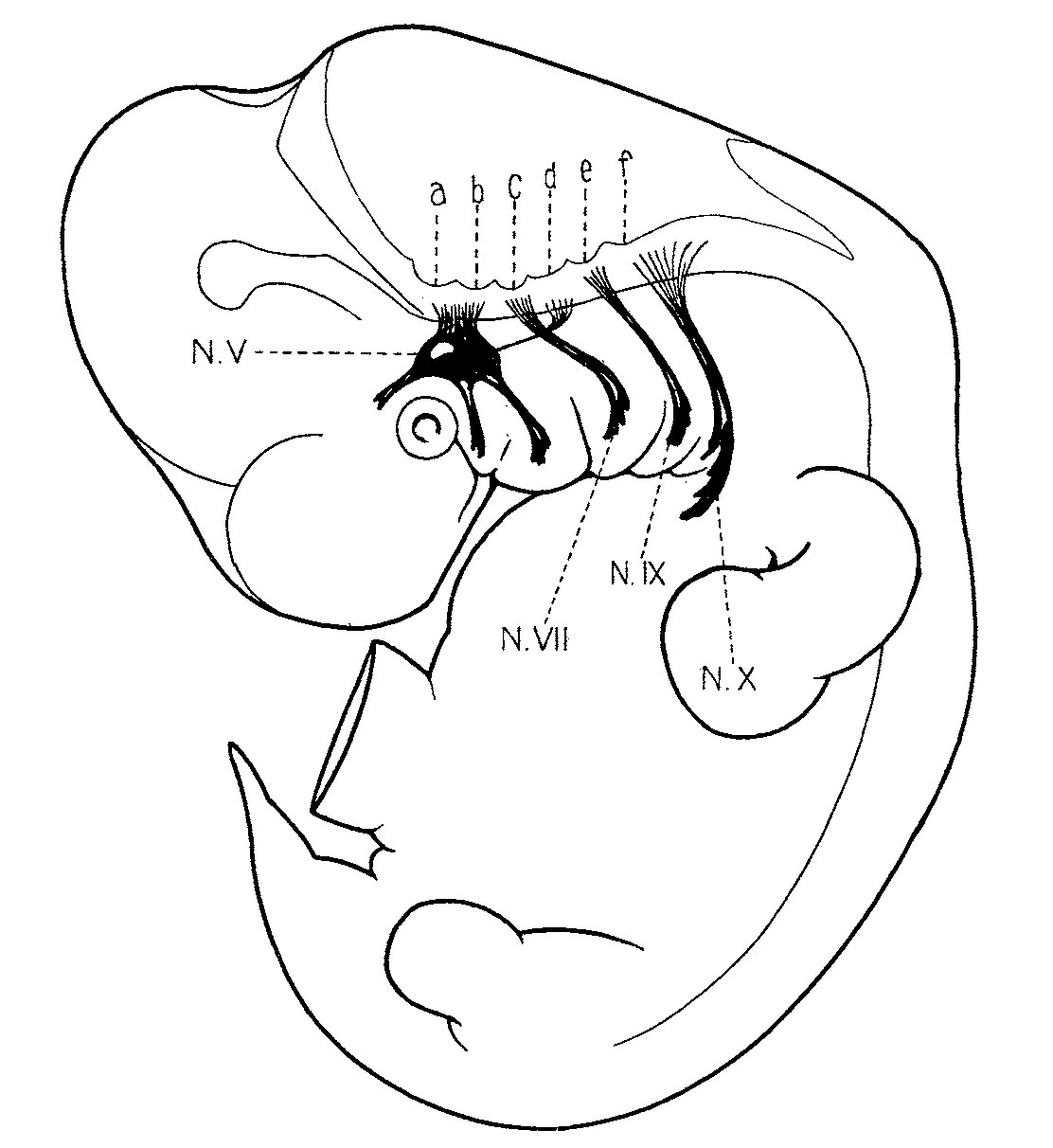 | 1,112 × 1,212 (132 KB) | Z8600021 (talk | contribs) | |
| 12:08, 14 September 2015 | 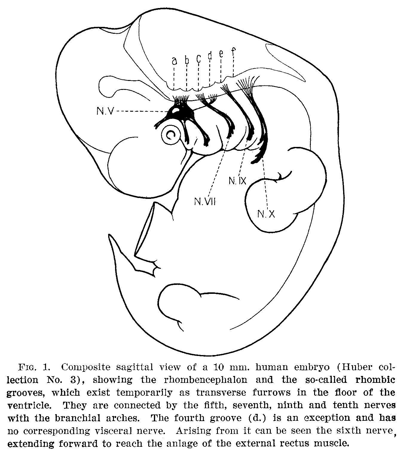 | 1,335 × 1,527 (263 KB) | Z8600021 (talk | contribs) |
You cannot overwrite this file.
File usage
The following 2 pages use this file: