Category:Computed Tomography: Difference between revisions
From Embryology
mNo edit summary |
mNo edit summary |
||
| (One intermediate revision by the same user not shown) | |||
| Line 1: | Line 1: | ||
This | This {{Embryology}} category shows media and pages related to the diagnostic tool of [[Computed Tomography]] imaging. | ||
===About Computed Tomography=== | |||
:'''Links:'''[[Computed Tomography]] | Computed Tomography or computed axial tomography (CAT or CT scan) began in 1970's using x-ray and a computer to produce images either as individual slices or reconstructed to give three dimensional (3D) views of specific anatomical regions or structures. Embryological applications include micro-CT (μCT) analysis. | ||
:'''Links:''' [[Computed Tomography]] | |||
Latest revision as of 11:42, 21 February 2015
This Embryology category shows media and pages related to the diagnostic tool of Computed Tomography imaging.
About Computed Tomography
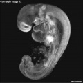
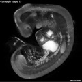
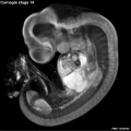
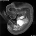
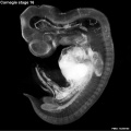
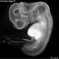
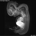
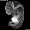
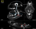
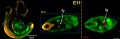
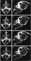
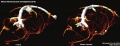
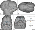
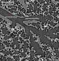
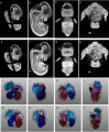
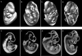
Computed Tomography or computed axial tomography (CAT or CT scan) began in 1970's using x-ray and a computer to produce images either as individual slices or reconstructed to give three dimensional (3D) views of specific anatomical regions or structures. Embryological applications include micro-CT (μCT) analysis.
- Links: Computed Tomography
Pages in category 'Computed Tomography'
The following 9 pages are in this category, out of 9 total.
Media in category 'Computed Tomography'
The following 90 files are in this category, out of 90 total.
 Absent cervical spine pedicle.jpg 600 × 338; 30 KB
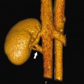
 Accessory renal artery.jpg 800 × 798; 103 KB
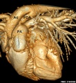
 Adult heart CT01.jpg 957 × 951; 212 KB
 Adult heart outflow tract CT01.jpg 747 × 747; 58 KB
 Adult heart outflow tract CT02.jpg 747 × 747; 65 KB
 Carnegie stage 12 OPT.jpg 800 × 801; 46 KB
 Carnegie stage 13 OPT.jpg 800 × 801; 53 KB
 Carnegie stage 14 OPT.jpg 800 × 801; 55 KB
 Carnegie stage 15 OPT.jpg 800 × 801; 56 KB
 Carnegie stage 16 OPT.jpg 800 × 801; 51 KB
 Carnegie stage 17 OPT.jpg 800 × 801; 57 KB
 Carnegie stage 18 OPT.jpg 800 × 801; 43 KB
 Carnegie stage 19 OPT.jpg 800 × 801; 46 KB
 Carnegie stage 20 OPT.jpg 800 × 801; 47 KB
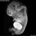
 Carnegie stage 21 OPT.jpg 800 × 801; 39 KB
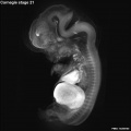
 Carnegie stage 22 OPT.jpg 800 × 801; 38 KB
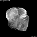
 Carnegie stage 23 OPT.jpg 800 × 801; 36 KB
 Cat inner ear MicroCT.jpg 1,159 × 1,300; 266 KB
 Choanal atresia computed tomography 01.jpg 598 × 477; 35 KB
 Cleidocranial dysplasia 01.jpg 518 × 700; 65 KB
 Computed tomography effective doses.jpg 600 × 441; 32 KB
 Dog patent ductus arteriosus computed tomography.jpg 600 × 654; 101 KB
 Ectopic pregnancy CT 01.jpg 800 × 676; 62 KB
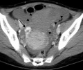 Ectopic pregnancy CT 02.jpg 800 × 676; 59 KB
 Ectopic pregnancy CT 03.jpg 800 × 676; 54 KB
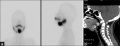 Ectopic thyroid - sublingual and suprahyoid.jpg 1,000 × 383; 42 KB
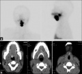 Ectopic thyroid - sublingual, suprahyoid and subhyoid.jpg 800 × 721; 59 KB
 Fetal mouth and palate day 64.jpg 1,890 × 2,126; 306 KB
 Fetus 35 week CT.jpg 700 × 874; 62 KB
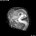
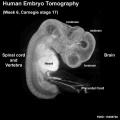 Human embryo tomography Carnegie stage 17.jpg 516 × 516; 35 KB
 Human inner ear MicroCT.jpg 2,131 × 3,111; 1,001 KB
 Human placenta vascular 01.jpg 1,200 × 644; 125 KB
 Human placenta vascular CT 01.jpg 938 × 1,000; 126 KB
 Hydatidiform mole pulmonary metastasis 01.jpg 1,000 × 750; 105 KB
 Incomplete cochlea CT.jpg 550 × 217; 24 KB
 Infant lymphocytic choriomeningitis virus CT.jpg 665 × 800; 69 KB
 Mouse CT axes E11.5.jpg 1,000 × 367; 68 KB
 Mouse CT E10.5 head 01.jpg 1,000 × 636; 154 KB
 Mouse CT E10.5 head.jpg 1,200 × 449; 83 KB
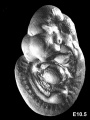 Mouse CT E10.5.jpg 602 × 800; 67 KB
 Mouse CT E11.5 movie-icon.jpg 335 × 375; 18 KB
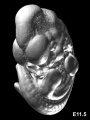 Mouse CT E11.5.jpg 602 × 800; 49 KB
 Mouse CT E12.5 scalebar.jpg 221 × 344; 4 KB
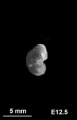
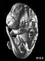 Mouse CT E12.5.jpg 602 × 800; 62 KB
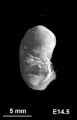 Mouse CT E14.5.jpg 221 × 344; 6 KB
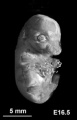 Mouse CT E16.5.jpg 221 × 344; 9 KB
 Mouse CT E18.5.jpg 221 × 344; 13 KB
 Mouse CT E9.5-E12 head.jpg 1,000 × 568; 56 KB
 Mouse E11 Foxf1.jpg 1,420 × 1,143; 162 KB
 Mouse E11 Sox2 and Nkx2.1.jpg 2,229 × 720; 176 KB
 Mouse E18 cerebral vasculature MicroCT.jpg 1,345 × 2,603; 411 KB
 Mouse E18 neurovasculature MicroCT.jpg 960 × 364; 68 KB
 Mouse E9 Foxf1.jpg 1,420 × 1,143; 142 KB
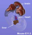
 Mouse embryo E11 and tomography 01.jpg 1,184 × 751; 120 KB
 Mouse embryo E11 HNF3beta notochord marker 01.jpg 2,245 × 829; 129 KB
 Mouse embryo E11 HNF3beta notochord marker 02.jpg 913 × 1,000; 68 KB
 Mouse embryo E11 HNF3beta notochord marker 03.jpg 913 × 1,000; 56 KB
 Mouse embryo E11 HNF3beta notochord marker 04.jpg 913 × 1,000; 55 KB
 Mouse embryo E11 tomography 01.jpg 628 × 800; 82 KB
 Mouse embryo E13 microCT icon.jpg 560 × 590; 35 KB
 Mouse embryo E14 microCT icon.jpg 858 × 720; 33 KB
 Mouse embryo E14 sectioned microCT icon.jpg 322 × 519; 31 KB
 Mouse embryo E15 microCT icon.jpg 347 × 490; 32 KB
 Mouse embryo E15 microCT.mov ; 3.71 MB
 Mouse embryo E15 microCT.mp4 ; 5.73 MB
 Mouse head E11.5 microCT 01.jpg 1,409 × 1,200; 308 KB
 Mouse lung micro-CT 01.jpg 650 × 668; 113 KB
 Mouse-E12.5.png 496 × 600; 278 KB
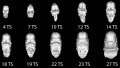
 Mouse-E9.5 E10.5 E11.5 E12.5.png 599 × 407; 170 KB
 Mouse-E9.5.jpg 602 × 800; 51 KB
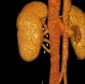
 Multiple renal arteries 01.jpg 496 × 496; 40 KB
 Postnatal persistant ductus venosus ultrasound 02.jpg 671 × 600; 44 KB
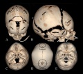
 Skull CT abnormal 01.jpg 1,000 × 549; 93 KB
 Skull CT abnormal 02.jpg 1,000 × 900; 119 KB
 Skull CT abnormal 03.jpg 1,000 × 542; 82 KB
 Skull CT abnormal 04.jpg 1,000 × 646; 102 KB
 Skull CT abnormal 05.jpg 1,000 × 572; 88 KB
 Skull CT abnormal 06.jpg 1,000 × 541; 85 KB
 Skull CT abnormal 07.jpg 1,000 × 541; 64 KB
 Skull CT abnormal 08.jpg 1,000 × 516; 73 KB
 Skull CT normal sutures 01.jpg 1,000 × 526; 89 KB
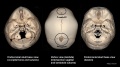 Skull CT normal sutures 02.jpg 1,000 × 559; 92 KB
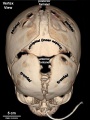 Skull CT normal sutures 03.jpg 600 × 800; 63 KB
 Skull CT normal sutures.jpg 1,000 × 900; 138 KB
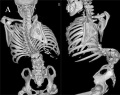 Spina bifida.jpg 800 × 633; 77 KB
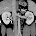
 Supernumerary renal vein 01.jpg 800 × 798; 72 KB
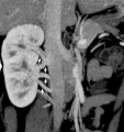
 Supernumerary renal vein 02.jpg 800 × 795; 89 KB
 Supernumerary renal vein 03.jpg 800 × 794; 80 KB
 Supernumerary renal vein 04.jpg 800 × 850; 76 KB


