Maternal-Fetal Medicine Trainees - Renal: Difference between revisions
mNo edit summary |
|||
| (6 intermediate revisions by the same user not shown) | |||
| Line 4: | Line 4: | ||
{| border='0px' | {| border='0px' | ||
|- | |- | ||
| | | width=420px|<html5media height="320" width="400">File:Nephron_development.mp4</html5media> | ||
[[ | [[Media:Nephron_development.mp4|'''Click Here''' to play on mobile device]] | ||
| valign="top" |'''Early Renal Development''' | | valign="top" |'''Early Renal Development''' | ||
| Line 17: | Line 17: | ||
'''Links:''' [[ | '''Links:''' [[Nephron Development Movie]] | [[Media:Nephron_development.mp4|MP4 version]] | [[Media:Nephron_development.mov|Quicktime version]] | [[Urogenital Sinus Movie]] | [[Renal System Development]] | ||
|- | |- | ||
|} | |} | ||
--[[User:Z8600021|Mark Hill]] ([[User talk:Z8600021|talk]]) 09:33, 14 May 2013 (EST) ''Please note, that software updates have led to movies requiring an update to MP4 versions to play on this current page. All functioning movies are available from [[Movies]] Page.'' | |||
==Textbooks== | ==Textbooks== | ||
{| | {| class="wikitable mw-collapsible mw-collapsed" | ||
| [ | ! References | ||
| | |- | ||
| {{Embryo logocitation}} | |||
The following chapter links only work with a UNSW connection | | | ||
{{Renal Links}} | |||
[http://php.med.unsw.edu.au/embryology/index.php?title=Lecture_-_Renal_Development&oldid=125206 2013] | [http://php.med.unsw.edu.au/embryology/index.php?title=Lecture_-_Renal_Development&oldid=108041 2012] | [http://php.med.unsw.edu.au/embryology/index.php?title=Lecture_-_Renal_Development&oldid=72417 2011] | |||
|- | |||
| {{MPT2011cover_citation}} | |||
| The following chapter links only work with a UNSW connection. | |||
* [http://er.library.unsw.edu.au/er/cgi-bin/eraccess.cgi?url=http://www.mdconsult.com/books/page.do?eid=4-u1.0-B978-1-4377-2002-0..00012-6&isbn=978-1-4377-2002-0&uniqId=330028653-2#4-u1.0-B978-1-4377-2002-0..00012-6 Chapter 12 - Urogenital System] | |||
|- | |- | ||
| | | {{SBBF2009cover_citation}} | ||
| | | The following chapter links only work with a UNSW connection. | ||
* [http://er.library.unsw.edu.au/er/cgi-bin/eraccess.cgi?url=http://www.mdconsult.com/books/linkTo?type=bookPage&isbn=978-0-443-06811-9&eid=4-u1.0-B978-0-443-06811-9..10015-6 Chapter 15 - Development of the Urogenital System] | |||
The following chapter links only work with a UNSW connection | |||
|- | |- | ||
| [[File: | | [[File:Endocrinology - An Integrated Approach.png|80px]] | ||
Nussey, S. and Whitehead, S. (2001). ''Endocrinology - An Integrated Approach''. UK Oxford: BIOS Scientific Publishers. ISBN-10: 1-85996-252-1 | |||
* | | [[Talk:Lecture_-_Endocrine_Development#Endocrinology_-_An_Integrated_Approach|Detailed Table of Contents]] | [http://www.ncbi.nlm.nih.gov/books/NBK22 Bookshelf Link] | ||
* [http://www.ncbi.nlm.nih.gov/books/n/endocrin/A972/ Chapter 6. The gonad] | |||
|} | |} | ||
| Line 66: | Line 71: | ||
===Kidney and Mesonephric Duct=== | ===Kidney and Mesonephric Duct=== | ||
{| | {| | ||
| | | width=380px|<html5media height="520" width="360">File:Urogenital_sinus_001.mp4</html5media> | ||
[[Media:Urogenital_sinus_001.mp4|'''Click Here''' to play on mobile device]] | |||
| '''First observe the development of the intermediate mesoderm.''' | | '''First observe the development of the intermediate mesoderm.''' | ||
| Line 103: | Line 109: | ||
{| border='0px' | {| border='0px' | ||
|- | |- | ||
| | | width=420px|<html5media height="320" width="400">File:Nephron_development.mp4</html5media> | ||
[[ | |||
[[Media:Nephron_development.mp4|'''Click Here''' to play on mobile device]] | |||
| valign="top" |'''Early Renal Development''' | | valign="top" |'''Early Renal Development''' | ||
| Line 121: | Line 128: | ||
'''Links:''' [[ | '''Links:''' [[Nephron Development Movie]] | [[Urogenital_Septum_Movie|Urogenital Sinus Movie]] | [[Renal System Development]] | ||
|- | |- | ||
|} | |} | ||
| Line 134: | Line 141: | ||
===Development of the Kidney=== | ===Development of the Kidney=== | ||
{| | {| | ||
| | | width=320px|<html5media height="460" width="300">File:Renal blood 01.mp4</html5media> | ||
[[Media:Renal blood 01.mp4|'''Click Here''' to play on mobile device]] | |||
| | | | ||
* starts in week 5 and is completed by week 15. | * starts in week 5 and is completed by week 15. | ||
| Line 145: | Line 154: | ||
===Development of the Urinary Bladder=== | ===Development of the Urinary Bladder=== | ||
{| | {| | ||
| | | width=460px|<html5media height="380" width="450">File:Trigone_001.mp4</html5media> | ||
[[Media:Trigone_001.mp4|'''Click Here''' to play on mobile device]] | |||
| '''Division of the Cloaca''' | | '''Division of the Cloaca''' | ||
Latest revision as of 11:11, 12 October 2014
Introduction

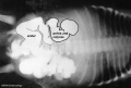
| <html5media height="320" width="400">File:Nephron_development.mp4</html5media> | Early Renal Development
This animation shows the process of early renal (kidney) development. Legend
|
--Mark Hill (talk) 09:33, 14 May 2013 (EST) Please note, that software updates have led to movies requiring an update to MP4 versions to play on this current page. All functioning movies are available from Movies Page.
Textbooks
| References | |
|---|---|
 |
|
 |
The following chapter links only work with a UNSW connection. |
 |
The following chapter links only work with a UNSW connection. |

Nussey, S. and Whitehead, S. (2001). Endocrinology - An Integrated Approach. UK Oxford: BIOS Scientific Publishers. ISBN-10: 1-85996-252-1 |
Detailed Table of Contents | Bookshelf Link |
- Links: More Embryology Textbooks
Renal Movies
|
|
|
| ||||||||||||
|
|
|
Urinary System Development


- The adult kidneys (the metanephroi) form from day 35, from a portion of the intermediate mesoderm called the metanephric blastema (or metanephric mesenchyme).
- They are induced to form by the ureteric buds, outgrowths from the end of the mesonephric ducts, which come into contact with the metanephric blastema.
- Upon contact, they begin to lengthen and bifurcate rapidly in the metanephric blastema – these branches differentiate into the collecting ducts.
- Both the ureteric buds and the metanephric blastema begin to differentiate; interestingly each induces differentiation in the other structure.
- The ureteric bud is induced by the metanephric blastema to form the collecting tubules, renal pelvis and ureters.
- The metanephric blastema is induced to form the nephrons.
- development occurs laterally symmetrical (left right)
- intermediate mesoderm lying beside the dorsal aorta
- initially form mesonephric tubules (epithelial)
- these tubules connect to a common duct, mesonephric duct
- the mesonephric duct then extends within the mesoderm, rostro-caudally
- eventually making contact with the cloaca
Kidney and Mesonephric Duct
| <html5media height="520" width="360">File:Urogenital_sinus_001.mp4</html5media> | First observe the development of the intermediate mesoderm.
|
Human Stages

|

|
| Embryo Stage 13 mesonephros (week 5) | Embryo Stage 22 metanephros (week 8) |

Kidney Nephron
Nephrogenesis is the process of generating new nephrons (continues to GA Week 36).
- 200,000 - 2,700,000.
- nephron number correlates with kidney volume.
| <html5media height="320" width="400">File:Nephron_development.mp4</html5media> | Early Renal Development
This animation shows the process of early renal (kidney) development. Legend
Sequence
|
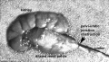
Adult Histology
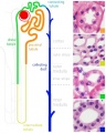
Adult nephron overview
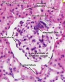
Glomerulus structure
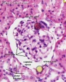
Vascular and renal poles


Development of the Kidney
| <html5media height="460" width="300">File:Renal blood 01.mp4</html5media> |
|
Development of the Urinary Bladder
| <html5media height="380" width="450">File:Trigone_001.mp4</html5media> | Division of the Cloaca
|
Development of the Urethra
- Further development of the urinary system varies depending on the sex of the embryo.
- Males - the pelvic urethra forms the membranous urethra, the prostatic urethra and penile urethra. (The sex of the above animation and sections is male)
- Females - the pelvic urethra forms the membranous urethra and the vestibule of the vagina.
Ten Most Frequently Reported Birth Anomalies
Based upon statistics from the Victorian Perinatal Data Collection Unit in Victoria (Australia) between 2003-2004.

|
Hypospadias (More? Development Animation - Genital Male External | Genital Abnormalities - Hypospadia) |

|
Obstructive Defects of the Renal Pelvis (obstructive defects of the renal pelvis, uteropelvic junction obstruction, pelvo-uterero junction obstruction) Term describing a developmental renal abnormality due to partial or complete blockage of the drainage of the kidney pelvis requiring surgical correction. The blockage can also have several causes including: unusual ureter twisting or bending, ureter compression by a blood vessel, malformations of the muscular wall. The blockage leads to an accumulation of urine in the affected region, with several potential effects: nephron damage from compression (hydronephrosis); decreased urine output leading to lack of amniotic fluid (oligohydramnios); respiratory development effects due to the lack of amniotic fluid.
(More? Renal System - Abnormalities | Renal System Development) |

|
Ventricular Septal Defect (More? Cardiovascular Abnormalities - Ventricular Septal Defect)
Heart Development Timeline (see Basic Cardiac Embryology) |

|
Congenital Dislocated Hip (More? Musculoskelal Abnormalities - Congenital Dislocation of the Hip (CDH))
(DHH, congenital dislocated hip, congenital hip dislocation, congenital hip dysplasia) Term describes a spectrum of musculoskeletal disorders of hip instability due either to the femoral head being able to move outside the acetabulum (luxation or dislocation), or abnormally within the acetabulum (subluxation or partial dislocation). This includes presentation following a normal examination of the hips in the newborn period (Ortolani and Barlow tests). When detected can be managed with splinting (Denis-Browne splint) allows the hip joint to develop normally and does not require surgery. If undetected and left untreated, the hip joint develops abnormally and surgical reduction is required. (More? Musculoskeletal System Development) |

|
Trisomy 21 or Down syndrome - (More? Trisomy 21) |

|
Hydrocephalus (More? Neural Abnormalities - Hydrocephalus |

|
Cleft Palate (More? Development Animation - Palate 1 | Development Animation - Palate 2 | Cleft Palate) |

|
Trisomy 18 or Edward Syndrome - multiple abnormalities of the heart, diaphragm, lungs, kidneys, ureters and palate 86% discontinued (More? Trisomy 18) |
| Renal Agenesis/Dysgenesis - reduction in neonatal death and stillbirth since 1993 may be due to the more severe cases being identified in utero and being represented amongst the increased proportion of terminations (approximately 31%). (More? Renal Abnormalities - Renal Agenesis) | |

|
Cleft Lip and Palate - occur with another defect in 33.7% of cases. (More? Cleft Lip) |

Abnormalities

Horseshoe Kidney

- fusion of the lower poles of the kidney.
- During migration from the sacral region the two metanephric blastemas can come into contact, mainly at the lower pole.
- The ureters pass in front of the zone of fusion of the kidneys.
- The kidneys and ureters usually function adequately but there is an increased incidence of upper urinary tract obstruction or infection.
- Some horseshoe variations have been described as having associated ureter abnormalities including duplications.
Urorectal Septum Malformation
- thought to be a deficiency in caudal mesoderm which in turn leads to the malformation of the urorectal septum and other structures in the pelvic region.
- Recent research has also identified the potential presence of a persistent urachus prior to septation of the cloaca (common urogenital sinus).
Bladder
- absent or small bladder -
associated with renal agenesis.
Bladder Exstrophy

- developmental abnormality associated with bladder development.
- origins appear to occur not just by abnormal bladder development, but by a congenital malformation of the ventral wall of abdomen (between umbilicus and pubic symphysis).
- There may also be other anomolies associated with failure of closure of abdominal wall and bladder (epispadias, pubic bone anomolies).
Ureter and Urethra
- Ureter - Duplex Ureter
- Urethra- Urethral Obstruction and Hypospadias
Polycystic Kidney Disease

- diffuse cystic malformation of both kidneys
- cystic malformations of liver and lung often associated, Often familial disposition
- Two types
- Infantile (inconsistent with prolonged survival)
- Adult (less severe and allows survival)
- Autosomal dominant PKD disease - recently identified at mutations in 2 different human genes encoding membrane proteins (possibly channels)
Wilms' Tumor
- (nephroblastoma) Named after Max Wilms, a German doctor who wrote first medical articles 1899
- most common type of kidney cancer children
- WT1 gene - encodes a zinc finger protein
- Both constitutional and somatic mutations disrupting the DNA-binding domain of WT1 result in a potentially dominant-negative phenotype
- some blastema cells (mass of undifferentiated cells) persist to form a ‘nephrogenic rest’
- Most rests become dormant or regress but others proliferate to form hyperplastic rests
- any type of rest can then undergo a genetic or epigenetic change to become a neoplastic rest
- can proliferate further to produce a benign lesion (adenomatous rest) or a malignant Wilms’ tumour
Obstructions
Hydronephrosis
Renal outflow obstruction
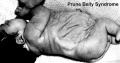
Prune_belly


- lower urinary tract obstruction
- mainly male
- fetal urinary system ruptures leading to collapse and "prune belly" appearance.
Glossary Links
- Glossary: A | B | C | D | E | F | G | H | I | J | K | L | M | N | O | P | Q | R | S | T | U | V | W | X | Y | Z | Numbers | Symbols | Term Link
Cite this page: Hill, M.A. (2024, June 14) Embryology Maternal-Fetal Medicine Trainees - Renal. Retrieved from https://embryology.med.unsw.edu.au/embryology/index.php/Maternal-Fetal_Medicine_Trainees_-_Renal
- © Dr Mark Hill 2024, UNSW Embryology ISBN: 978 0 7334 2609 4 - UNSW CRICOS Provider Code No. 00098G