File:Early zygote labelled.jpg: Difference between revisions
mNo edit summary |
m (→Early Zygote) |
||
| Line 13: | Line 13: | ||
* the zygote floats freely within the uterine tube. | * the zygote floats freely within the uterine tube. | ||
* The cell is preparing for the first mitotic division. | * The cell is preparing for the first mitotic division. | ||
{{Zygote image links}} | {{Zygote image links}} | ||
Revision as of 19:24, 6 April 2014
Early Zygote
This is described as an early human zygote due to the presence of the 2 pronuclei (male and female) in the centre of the cytoplasm.
The haploid pronuclei of the male (spermatozoa) and female (oocyte) have not yet combined to form a single nucleus.
The polar bodies can be seen at the edge of the cytoplasm (at 3 o'clock position). These exclusion bodies contain the additional oocyte DNA produced in meiosis.
The zona pellucida forms the thick clear layer that surrounds the cell.
At this stage in vivo:
- there would still be granulosa cells and spermatozoa attached to the zone pellucida.
- the zygote floats freely within the uterine tube.
- The cell is preparing for the first mitotic division.
- Zygote Image Links: Image - Early zygote | Image - Early zygote labelled | Image - zygote 1 | Image - zygote 2 | Image - zygote 3 | Image - zygote 2 labeled | Fertilization | Zygote | Carnegie stage 1 | Category:Zygote
About Carnegie Stages 1
Facts: Week 1, size 0.1 - 0.15 mm (100 - 150 microns)
Features: zygote, fertilized oocyte, pronuclei, polar bodies, zona pellucida
Image Source: UNSW Embryology http://embryology.med.unsw.edu.au/wwwhuman/Stages/Stage1.htm
Cite this page: Hill, M.A. (2024, June 14) Embryology Early zygote labelled.jpg. Retrieved from https://embryology.med.unsw.edu.au/embryology/index.php/File:Early_zygote_labelled.jpg
- © Dr Mark Hill 2024, UNSW Embryology ISBN: 978 0 7334 2609 4 - UNSW CRICOS Provider Code No. 00098G
File history
Click on a date/time to view the file as it appeared at that time.
| Date/Time | Thumbnail | Dimensions | User | Comment | |
|---|---|---|---|---|---|
| current | 11:47, 10 March 2012 | 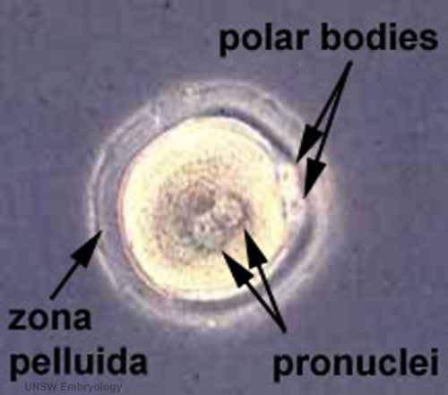 | 500 × 441 (29 KB) | Z8600021 (talk | contribs) | increase image size |
| 13:28, 20 July 2010 |  | 216 × 191 (8 KB) | S8600021 (talk | contribs) | ==Early Zygote (labelled)== About Carnegie Stages 1 Facts: Week 1, size 0.1-0.15 mm Features: zygote, fertilized oocyte, pronuclei, polar bodies, zona pellucida Related Images: Early zygote | Image Source: UNSW Embryology ht |
You cannot overwrite this file.
File usage
The following 14 pages use this file:
- 2015 Group Project 1
- BGDA Lecture - Development of the Embryo/Fetus 1
- Carnegie stage 1
- Embryonic Development
- Fertilization
- Lecture - 2015 Course Introduction
- Lecture - 2016 Course Introduction
- Lecture - 2017 Course Introduction
- Lecture - Fertilization
- P
- Z
- Zygote
- Talk:2015 Group Project 1
- Talk:Lecture - 2016 Course Introduction