File:Eye and retina cartoon.jpg: Difference between revisions
mNo edit summary |
|||
| (9 intermediate revisions by the same user not shown) | |||
| Line 4: | Line 4: | ||
| valign="top" width="250px"|'''The Eye''' | | valign="top" width="250px"|'''The Eye''' | ||
* Retina forms the inner lining of the most of the posterior part of the eye. | * Retina forms the inner lining of the most of the posterior part of the eye. | ||
* The RPE is sandwiched between the retina and choroids, a vascularized and pigmented connective tissue. | * The RPE is sandwiched between the retina and choroids, a vascularized and pigmented connective tissue. | ||
* An enlarged diagram of the fovea is shown in the box. | |||
| valign="top" width=" | | valign="top" width="200px"|'''Retinal Cell Organization''' | ||
* '''G''' - ganglion cells | * '''G''' - retinal ganglion cells | ||
* '''M''' - Müller cell | * '''M''' - Müller cell | ||
* '''A''' - amacrine cell | * '''A''' - amacrine cell | ||
| Line 29: | Line 29: | ||
Retina | :'''Links:''' [[Sensory - Vision Development|Vision Development]] | [[Vision - Retina Development|Retina Development]] | ||
| Line 35: | Line 35: | ||
<pubmed>20855501</pubmed>| [http://www.ncbi.nlm.nih.gov/pmc/articles/PMC3101587 PMC3101587] | [http://jcb.rupress.org/content/190/6/953.full JCB] | <pubmed>20855501</pubmed>| [http://www.ncbi.nlm.nih.gov/pmc/articles/PMC3101587 PMC3101587] | [http://jcb.rupress.org/content/190/6/953.full JCB] | ||
[[:File:Human-retina-01.jpg|Retina histology image]] - Swaroop and Zack (2002), published by BioMed Central PMID 12186651 | |||
{{JCB}} | |||
Note - '''Retinal Cell Organization''' and '''Human Retina Histology''' were flipped vertically and relabelled from original published image to match the orientation in '''The Eye'''. This is the ongoing issue of which way around the retina should be illustrated. For an introduction to this topic it is just easier to have it the same way around. | |||
---- | |||
{{Footer}} | |||
[[Category:Vision]] [[Category:Cartoon]] | [[Category:Vision]] [[Category:Cartoon]] | ||
Latest revision as of 04:58, 4 January 2014
Eye and Retina
The Eye
|
Retinal Cell Organization
|
Human Retina Histology
|
- Links: Vision Development | Retina Development
Reference
<pubmed>20855501</pubmed>| PMC3101587 | JCB
Retina histology image - Swaroop and Zack (2002), published by BioMed Central PMID 12186651
Copyright
Rockefeller University Press - Copyright Policy This article is distributed under the terms of an Attribution–Noncommercial–Share Alike–No Mirror Sites license for the first six months after the publication date (see http://www.jcb.org/misc/terms.shtml). After six months it is available under a Creative Commons License (Attribution–Noncommercial–Share Alike 4.0 Unported license, as described at https://creativecommons.org/licenses/by-nc-sa/4.0/ ). (More? Help:Copyright Tutorial)
Note - Retinal Cell Organization and Human Retina Histology were flipped vertically and relabelled from original published image to match the orientation in The Eye. This is the ongoing issue of which way around the retina should be illustrated. For an introduction to this topic it is just easier to have it the same way around.
Cite this page: Hill, M.A. (2024, May 31) Embryology Eye and retina cartoon.jpg. Retrieved from https://embryology.med.unsw.edu.au/embryology/index.php/File:Eye_and_retina_cartoon.jpg
- © Dr Mark Hill 2024, UNSW Embryology ISBN: 978 0 7334 2609 4 - UNSW CRICOS Provider Code No. 00098G
File history
Click on a date/time to view the file as it appeared at that time.
| Date/Time | Thumbnail | Dimensions | User | Comment | |
|---|---|---|---|---|---|
| current | 14:50, 4 March 2012 | 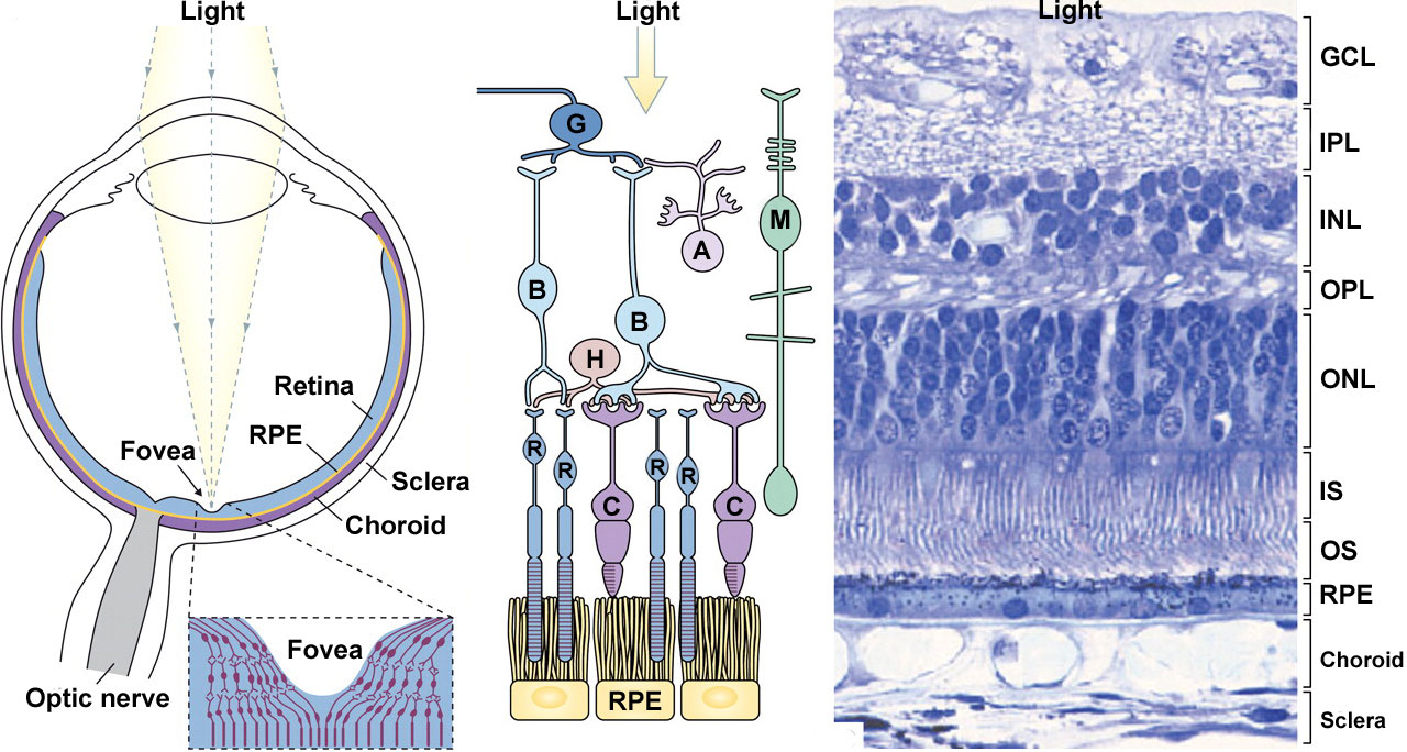 | 1,280 × 684 (211 KB) | Z8600021 (talk | contribs) |
You cannot overwrite this file.